

Identification of two functional nuclear localization signals mediating nuclear import of liver receptor homologue-1
- PMID: 20853131
- PMCID: PMC11114538
- DOI: 10.1007/s00018-010-0521-5
Identification of two functional nuclear localization signals mediating nuclear import of liver receptor homologue-1
Abstract
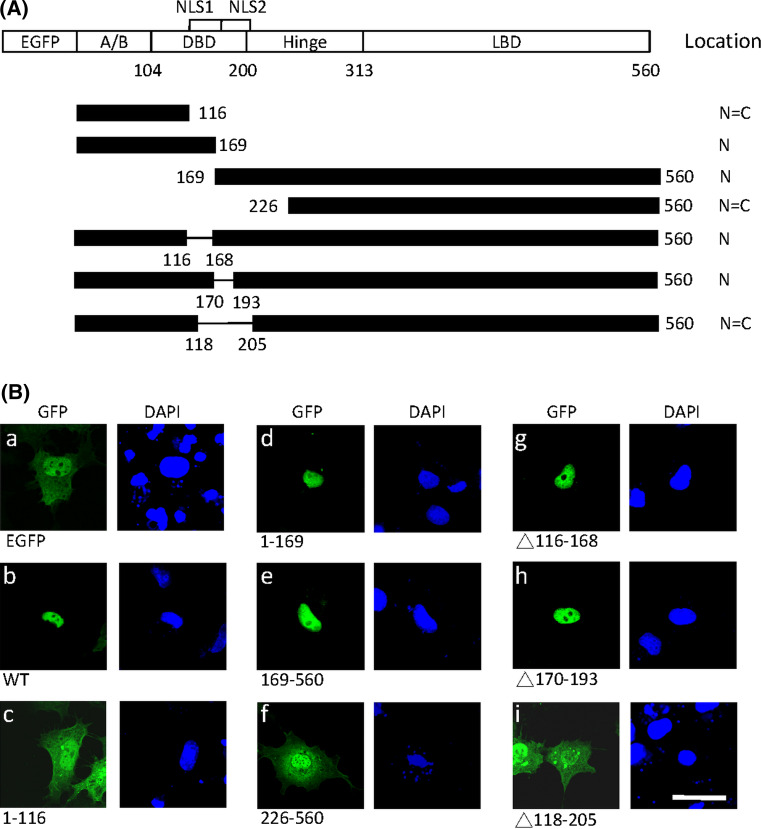
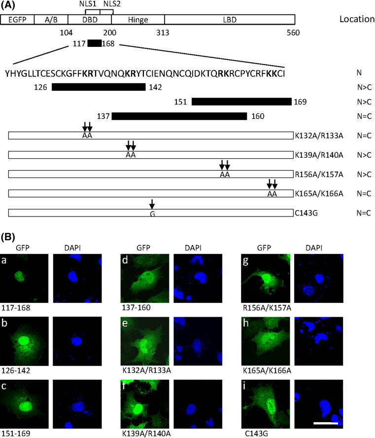
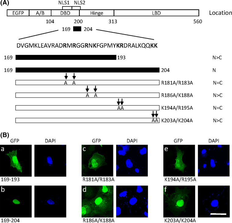
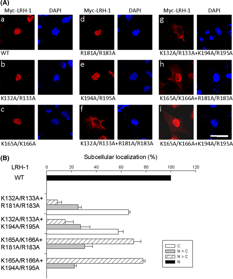
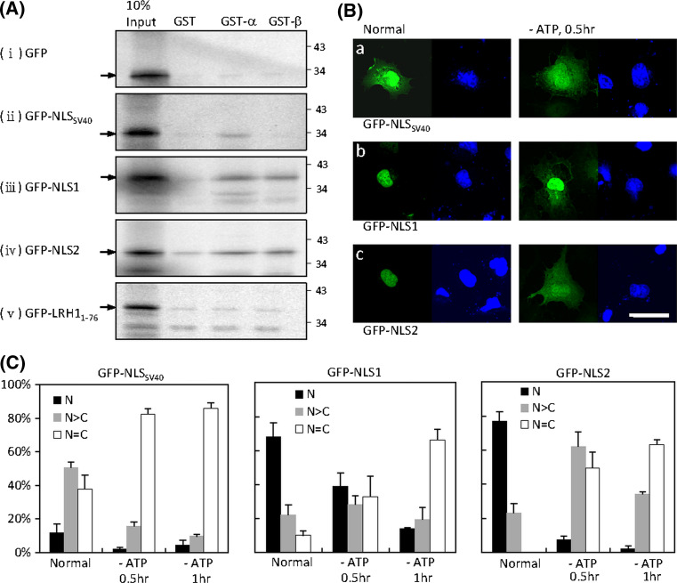
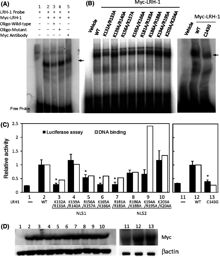
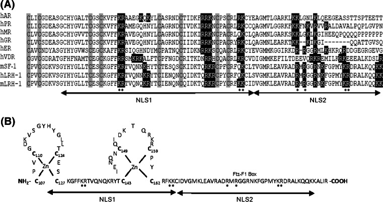
Liver receptor homologue-1 (LRH-1) is a member of the nuclear receptor superfamily. We characterized two functional nuclear localization signals (NLSs) in LRH-1. NLS1 (residues 117-168) overlaps the second zinc finger in the DNA binding domain. Mutagenesis showed that the zinc finger structure and two basic clusters on either side of the zinc finger loop are critical for nuclear import of NLS1. NLS2 (residues 169-204) is located in the Ftz-F1 box that contains a bipartite signal. In full-length LRH-1, mutation of either NLS1 or NLS2 had no effect on nuclear localization, but disruption of both NLS1 and NLS2 resulted in the cytoplasmic accumulation of LRH-1. Either NLS1 or NLS2 alone was sufficient to target LRH-1 to the nucleus. Both NLS1 and NLS2 mediate nuclear transport by a mechanism involving importin α/β. Finally, we showed that three crucial basic clusters in the NLSs are involved in the DNA binding and transcriptional activities of LRH-1.
Figures
Similar articles
-
Identification of nuclear import and export signals within Fli-1: roles of the nuclear import signals in Fli-1-dependent activation of megakaryocyte-specific promoters.Mol Cell Biol. 2005 Apr;25(8):3087-108. doi: 10.1128/MCB.25.8.3087-3108.2005. Mol Cell Biol. 2005. PMID: 15798196 Free PMC article.
-
Daxx contains two nuclear localization signals and interacts with importin alpha3.J Cell Biochem. 2008 Feb 1;103(2):456-70. doi: 10.1002/jcb.21408. J Cell Biochem. 2008. PMID: 17661348
-
An importin alpha/beta-recognized bipartite nuclear localization signal mediates targeting of the human herpes simplex virus type 1 DNA polymerase catalytic subunit pUL30 to the nucleus.Biochemistry. 2007 Aug 14;46(32):9155-63. doi: 10.1021/bi7002394. Epub 2007 Jul 20. Biochemistry. 2007. PMID: 17640102
-
Nuclear import of aristaless-related homeobox protein via its NLS1 regulates its transcriptional function.Mol Cell Biochem. 2013 Sep;381(1-2):221-31. doi: 10.1007/s11010-013-1706-7. Epub 2013 Jun 16. Mol Cell Biochem. 2013. PMID: 23771350
-
Identification and characterization of two putative nuclear localization signals (NLS) in the DNA-binding protein NUCKS.Biochim Biophys Acta. 2007 Sep;1773(9):1398-406. doi: 10.1016/j.bbamcr.2007.05.013. Epub 2007 Jun 2. Biochim Biophys Acta. 2007. PMID: 17604136
Cited by
-
Regulation of TLR4 signaling through the TRAF6/sNASP axis by reversible phosphorylation mediated by CK2 and PP4.Proc Natl Acad Sci U S A. 2021 Nov 23;118(47):e2107044118. doi: 10.1073/pnas.2107044118. Proc Natl Acad Sci U S A. 2021. PMID: 34789577 Free PMC article.
-
Nuclear pore complex evolution: a trypanosome Mlp analogue functions in chromosomal segregation but lacks transcriptional barrier activity.Mol Biol Cell. 2014 May;25(9):1421-36. doi: 10.1091/mbc.E13-12-0750. Epub 2014 Mar 5. Mol Biol Cell. 2014. PMID: 24600046 Free PMC article.
-
sNASP inhibits TLR signaling to regulate immune response in sepsis.J Clin Invest. 2018 Jun 1;128(6):2459-2472. doi: 10.1172/JCI95720. Epub 2018 May 7. J Clin Invest. 2018. PMID: 29733298 Free PMC article.
-
PEP-sNASP Peptide Alleviates LPS-Induced Acute Lung Injury Through the TLR4/TRAF6 Axis.Front Med (Lausanne). 2022 Mar 21;9:832713. doi: 10.3389/fmed.2022.832713. eCollection 2022. Front Med (Lausanne). 2022. PMID: 35386914 Free PMC article.
-
Strategies on the nuclear-targeted delivery of genes.J Drug Target. 2013 Dec;21(10):926-39. doi: 10.3109/1061186X.2013.830310. Epub 2013 Aug 22. J Drug Target. 2013. PMID: 23964565 Free PMC article. Review.
References
-
- Boerboom D, Pilon N, Behdjani R, Silversides DW, Sirois J. Expression and regulation of transcripts encoding two members of the NR5A nuclear receptor subfamily of orphan nuclear receptors, steroidogenic factor-1 and NR5A2, in equine ovarian cells during the ovulatory process. Endocrinology. 2000;141:4647–4656. doi: 10.1210/en.141.12.4647. - DOI - PubMed
-
- Pare JF, Malenfant D, Courtemanche C, Jacob-Wagner M, Roy S, Allard D, Belanger L. The fetoprotein transcription factor (FTF) gene is essential to embryogenesis and cholesterol homeostasis and is regulated by a DR4 element. J Biol Chem. 2004;279:21206–21216. doi: 10.1074/jbc.M401523200. - DOI - PubMed
-
- Morohashi KI, Omura T. Ad4BP/SF-1, a transcription factor essential for the transcription of steroidogenic cytochrome P450 genes and for the establishment of the reproductive function. FASEB J. 1996;10:1569–1577. - PubMed
Publication types
MeSH terms
Substances
LinkOut - more resources
Full Text Sources
