Review
. 2010 Nov;20(6):380-91.
doi: 10.1002/rmv.666.

Structure of influenza virus ribonucleoprotein complexes and their packaging into virions

Affiliations
Review

Structure of influenza virus ribonucleoprotein complexes and their packaging into virions

Takeshi Noda et al. Rev Med Virol. 2010 Nov.

Abstract

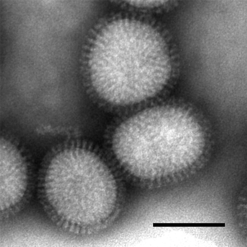
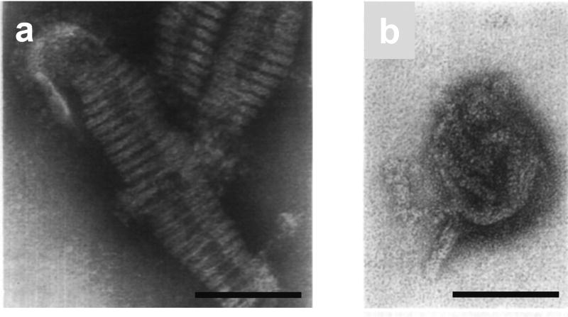
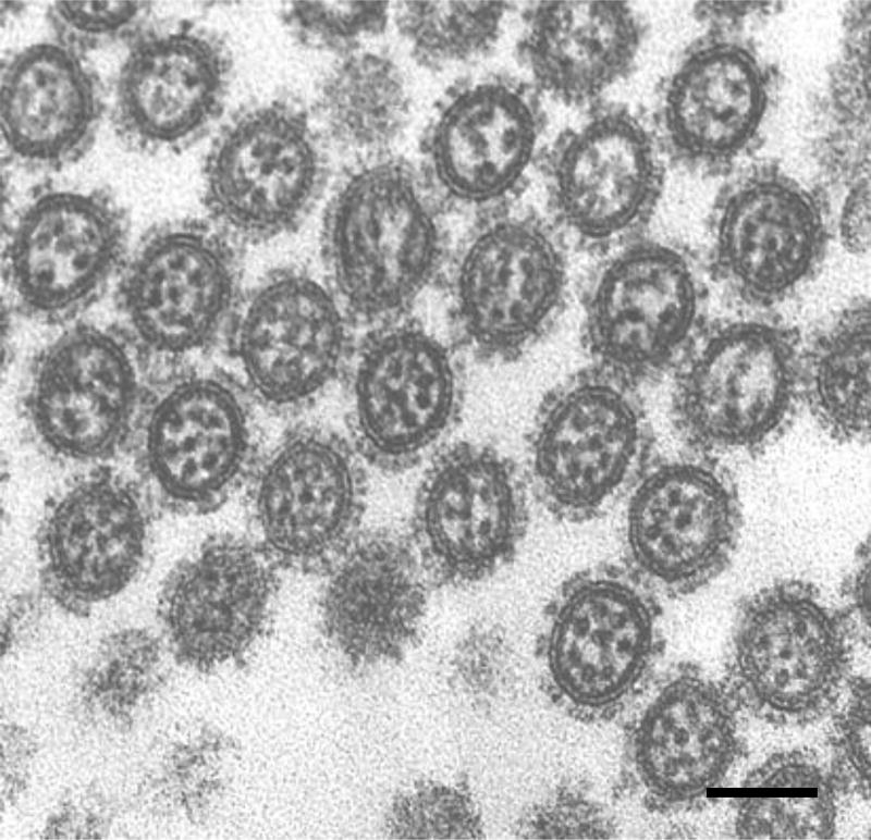
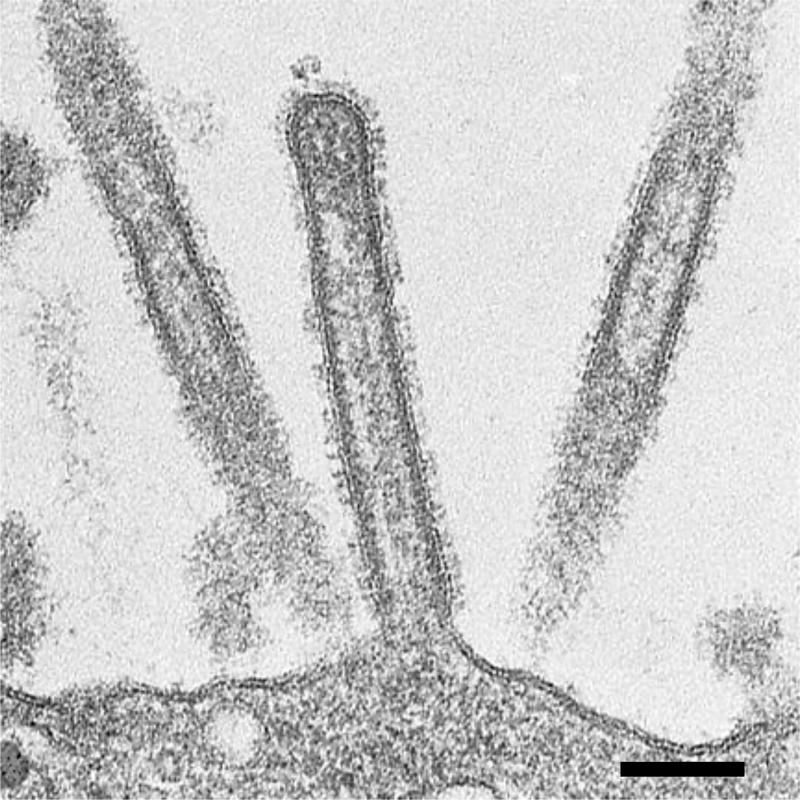
The influenza A virus genome consists of eight segmented, single-stranded, negative-sense RNAs. Each viral RNA (vRNA) segment forms a ribonucleoprotein (RNP) complex together with NPs and a polymerase complex, which is a fundamental unit for transcription and replication of the viral genome. Although the exact structure of the intact RNP remains poorly understood, recent electron microscopic studies have revealed certain structural characteristics of the RNP. This review focuses on the findings of these various electron microscopic analyses of RNPs extracted from virions and RNPs inside virions. Based on the morphological and structural observations, we present the architecture of RNPs within a virion and discuss the genome packaging mechanism by which the vRNA segments are incorporated into virions.


Figures

Figure 1
Figure 1
Purified influenza virions visualized by negative staining electron microscopy. The spherical virions, approximately 120 nm in diameter, are covered with spikes composed of HA and NA. Bar; 100 nm.
Figure 2
Figure 2
Schematic diagram of an extracted RNP. The RNP consists of a strand of NP monomers folded back on itself creating a loop at the end, and coiled on itself, forming a double-stranded helical body except at the loop. The NP monomers and vRNA are depicted as gray spheres and a blue ribbon, respectively The polymerase complex is depicted by a yellow box, a red cone, and a green rod.
Figure 3
Figure 3
Internal structure of purified virions observed by negative staining electron microscopy. (a) A continuous helix approximately 8 nm in diameter is observed in a disrupted virion. (b) Irregularly compressed rod-like structures approximately 12 nm in diameter. Two rod-like structures can be seen protruding from the virion. These electron micrographs have been reproduced with permission from those published in Schulze (1972) (54). Bars; 100 nm.
Figure 4
Figure 4
Internal structure of budding virions revealed by thin-section electron microscopy. Transversely sectioned budding virions show a well-organized arrangement of eight RNPs within the virions. Some virions contain less than eight RNPs because the RNPs differ in length. Thus, the number of RNPs seen in the virions varies depending on where the budding virions are transversely sectioned. Bar; 100 nm.
Figure 5
Figure 5
Longitudinal section of filamentous virions budding from the cell surface. The rod-like RNPs are associated with the envelope at the distal end of the budding virion. Note that the RNPs are present only at the distal end of the virion. Bar; 100 nm.
Figure 6
Figure 6
Schematic diagram of RNPs within a budding virion. (a–c) Incorporation of the eight RNPs is coordinated with bud growth. In an elongated budding virion, eight RNPs are arranged into the (7+1) configuration. (d) In a spherical virion released from the plasma membrane, the RNPs are compressed and the arrangement of eight RNPs is disordered.

Similar articles

Cited by

References

    1. Palese P. Orthomyxoviridae. In: Knipe DM, Howley PM, Griffin DE, Lamb RA, Martin MA, Roizman B, Straus SE, editors. Fields virology. 5. Lippincott Williams & Wilkins; Philadelphia: 2007. pp. 1647–1689.
    1. Fouchier RA, Munster V, Wallensten A, et al. Characterization of a novel influenza A virus hemagglutinin subtype (H16) obtained from black-headed gulls. J Virol. 2005;79:2814–2822. - PMC - PubMed
    1. Osterhaus AD, Rimmelzwaan GF, Martina BE, et al. Influenza B virus in seals. Science. 2000;288:1051–1053. - PubMed
    1. Guo YJ, Jin FG, Wang P, et al. Isolation of influenz C virus from pigs and experimental infection of pigs with influenza C virus. J Gen Virol. 1983;64:177–182. - PubMed
    1. Taylor RM. Studies on survival of influenza virus between epidemics and antigenic variants of the virus. Am J Public Health Nations Health. 1949;39:171–178. - PMC - PubMed

Publication types

MeSH terms

Substances