

Pleistophora hyphessobryconis (Microsporidia) infecting zebrafish Danio rerio in research facilities
- PMID: 20853741
- PMCID: PMC4155925
- DOI: 10.3354/dao02245
Pleistophora hyphessobryconis (Microsporidia) infecting zebrafish Danio rerio in research facilities
Abstract
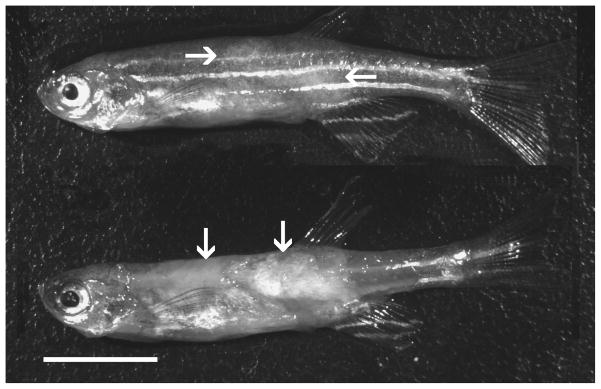
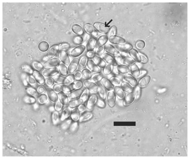
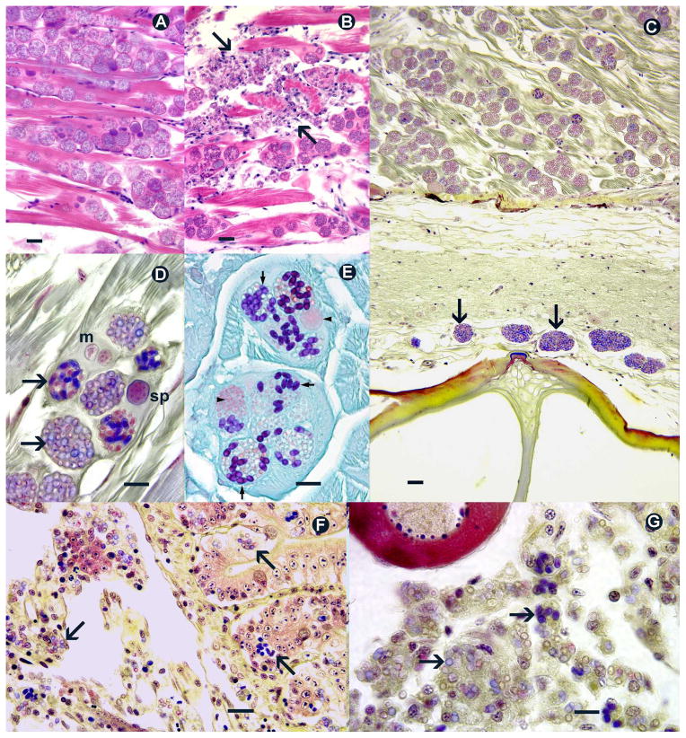
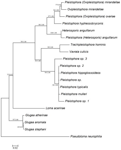
Zebrafish Danio rerio are important models for biomedical research, and thus, there is an increased concern about diseases afflicting them. Here we describe infections by Pleistophora hyphessobryconis (Microsporidia) in zebrafish from 3 laboratories. As reported in other aquarium fishes, affected zebrafish exhibited massive infections in the skeletal muscle, with no involvement of smooth or cardiac muscle. In addition, numerous spores within macrophages were observed in the visceral organs, including the ovaries. Transmission studies and ribosomal RNA (rRNA) gene sequence comparisons confirmed that the parasite from zebrafish was P. hyphessobryconis as described from neon tetra Paracheirodon innesi. Ten 15 d old zebrafish were exposed to P. hyphessobryconis collected from 1 infected neon tetra, and 7 of 10 fish became infected. Comparison of P. hyphessobryconis small subunit rRNA gene sequence from neon tetra with that obtained from zebrafish was nearly identical, with < 1% difference. Given the severity of infections, P. hyphessobryconis should be added to the list of pathogens that should be avoided in zebrafish research facilities, and it would be prudent to avoid mixing zebrafish used in research with other aquarium fishes.
Figures
References
-
- Ackermann G. Zebrafish: a genetic model for vertebrate organogenesis and human disorders. Front Biosci. 2003;8:d1227–d1253. - PubMed
-
- Altschul SF, Gish W, Miller W, Myers EW, Lipman DJ. Basic local alignment search tool. J Mol Biol. 1990;215:403–410. - PubMed
-
- Amatruda JF, Shepard JL, Stern HM, Zon LI. Zebrafish as a cancer model system. Cancer Cell. 2002;1:229–231. - PubMed
-
- Bruno DW, Nowak B, Elliott DG. Guide to the identification of fish protozoan and metazoan parasites in stained tissue sections. Dis Aquat Org. 2006;70:1–36. - PubMed
-
- Canning EU, Lom J, Dyková I. The Microsporidia of Vertebrates. London: Academic Press; 1986. Description of species infecting fish: Pleistophora hyphessobryconis Schäperclaus, 1941; pp. 101–107.
Publication types
MeSH terms
Substances
Grants and funding
LinkOut - more resources
Full Text Sources
Research Materials
