. 2011 Jun;20(6):1021-9.
doi: 10.1089/scd.2010.0198. Epub 2010 Nov 9.

SDF-1α secreted by human CD133-derived multipotent stromal cells promotes neural progenitor cell survival through CXCR7

Affiliations

SDF-1α secreted by human CD133-derived multipotent stromal cells promotes neural progenitor cell survival through CXCR7

Benjamin Bakondi et al. Stem Cells Dev. 2011 Jun.

Abstract

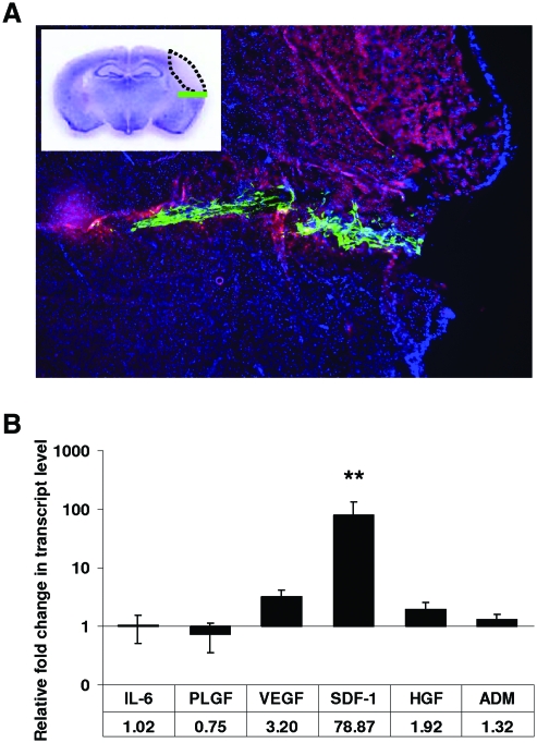
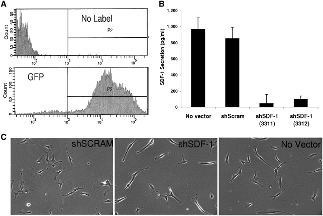
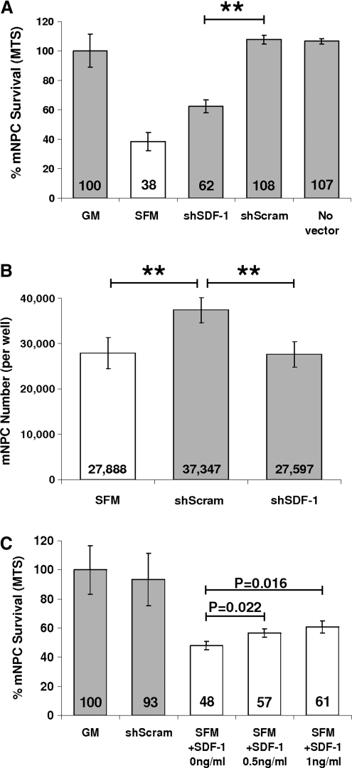
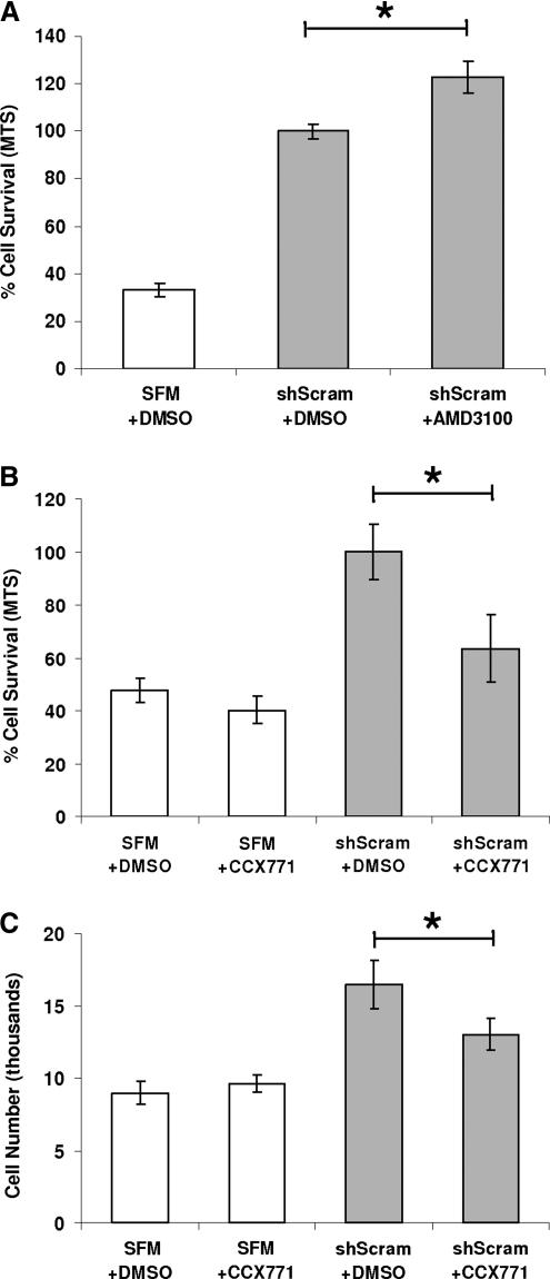
We recently reported that concentrated conditioned medium (CdM) from human CD133-derived bone marrow progenitor cells (CD133 CdM) was neuroprotective after stroke. Here we identify stromal-derived factor 1 alpha (SDF-1) as a potential neuroprotective candidate in CD133 CdM by interrogating the transcriptional responses of CD133-derived multipotent stromal cells (CD133dMSCs) after cell injection into the ischemic brain. Human SDF-1 mRNA was upregulated 79-fold by CD133dMSCs when injected into the stroke peri-infarct area compared with cells injected into the uninjured parenchyma of sham-operated animals. In cell protection assays, we replaced the typical growth medium in mouse neural progenitor cell (mNPC) cultures with serum-free CD133 CdM immediately before exposure to hypoxia (1% oxygen) for 48 h. CD133 CdM significantly increased the survival of mNPCs during hypoxia exposure and growth factor withdrawal. To determine whether MSC-secreted SDF-1 influenced mNPC survival, we used lentiviral short hairpin RNA against SDF1 (shSDF-1) to knockdown SDF-1 expression in CD133dMSCs. The CdM generated from shSDF-1-treated cells had a 94% decrease in secreted SDF-1 and was significantly less protective for mNPCs when compared with control CdM from CD133dMSCs transduced with scrambled short hairpin RNA. Pharmacological inhibition of the 2 known SDF-1 receptors, CXCR4 and CXCR7, revealed that only CXCR7 activity was functionally linked to survival signaling in mNPCs during hypoxia exposure. Treatment of mNPCs with CD133 CdM and CXCR7 inhibitor decreased mNPC viability by 36.5% ± 12.8% and decreased cell number by 21% ± 6.7% compared with dimethyl sulfoxide treated controls. These data indicate that SDF-1 is a key neuroprotective cytokine secreted by CD133dMSCs that protects mNPCs through CXCR7.


Figures

FIG. 1.
FIG. 1.
CD133-derived multipotent stromal cells (CD133dMSCs) increased their stromal-derived factor 1 alpha (SDF-1) mRNA levels at the stroke peri-infarct area. (A) Epifluorescence image of coronal section from a representative mouse brain that underwent middle cerebral artery ligation (MCAL) and was injected with 1.25 × 105 green fluorescent protein (GFP) CD133dMSCs the following day. Red autofluorescence indicates the stroke area and GFP CD133dMSCs localize to the needle track in the peri-infarct area 48 h postinjection. Inset: cresyl violet stain and schematic of stroke area (dotted black line) and injected cells (green line). (B) Human-specific quantitative real time (RT)-polymerase chain reaction for mRNAs of CD133dMSC secreted-factors was performed on RNA isolated from brain tissues of mice that underwent MCAL or sham surgery 3 days prior. Numerical values indicate the fold change in mRNA levels for each factor in MCAL versus sham-operated mice. The mRNA level for each sample was normalized to its corresponding human glyceraldehyde 3-phosphate dehydrogenase mRNA level. mRNA levels were quantified by the ΔΔCt method. Animal numbers for each factor: interleukin 6 (IL-6): 4 sham and 4 stroke; adrenomedullin (ADM): 8 sham and 8 stroke; vascular endothelial growth factor (VEGF): 5 sham and 8 stroke; SDF-1: 10 sham and 12 stroke; placental growth factor (PLGF): 4 sham and 4 stroke; hepatocyte growth factor (HGF): 7 sham and 6 stroke. **P ≤ 0.01. Error bars indicate standard error of mean. Color images available online at www.liebertonline.com/scd
FIG. 2.
FIG. 2.
Expression of CXCR4 and CXCR7 by mouse neural progenitor cells (mNPCs). (A) mNPCs are multipotent. Triple staining is shown for astrocytes (GFAP, blue), neurons (Beta-III tubulin, red), and oligodendrocytes (O4, green). (B) Floating neural spheres before trituration to single cell suspension. (C) After trituration, the single cell suspension was plated and expanded as NPCs. (D) RT-PCR for mRNAs of CXCR4, CXCR7, and glyceraldehyde 3-phosphate dehydrogenase. Bottom panels show no reverse transcriptase controls for each primer set. (E–G) Immunocytochemistry for CXCR4. (H–J) Isotype controls for CXCR4 (rabbit isotype). (K–M) Immunocytochemistry for CXCR7. (N–P) Isotype controls for CXCR7 (mouse isotype). Insets: close-up views of cell staining. Color images available online at www.liebertonline.com/scd
FIG. 3.
FIG. 3.
Knockdown of SDF-1 secretion in CD133dMSCs by lentiviral short hairpin RNA (shRNA). CD133dMSCs were transduced with puromycin-selectable lentivectors expressing GFP (transduction control), scrambled (nonspecific) shRNA, or sequence-specific shRNA (against SDF-1). (A) Fluorescent activated cell sorting (FACS) of control cells (no label) and those transduced with GFP lentivector and selected by puromycin to determine cell purity after selection regime. (B) Enzyme-linked immunosorbent assay for SDF-1 showed a 94% and 89% reduction in level of secreted SDF-1 from CD133dMSCs after lentiviral transduction of 2 different shRNA sequences for the SDF-1 transcript compared with a scrambled vector. SDF-1 secretion levels were not different from cells that received the scrambled vector and cells that were not transduced (no vector control). All measurements were performed in triplicate. (C) Phase-contrast microscopy images of CD133dMSCs that received lentivectors and no vector control cells (magnification 10×). shSDF-1, shRNA specific to SDF-1; shScram, scrambled non-specific shRNA (control).
FIG. 4.
FIG. 4.
SDF-1 knockdown diminishes the protective effect of the conditioned medium (CdM). (A, B) mNPC survival was significantly reduced after 48 h of hypoxic culture when treated with shSDF-1 CdM compared with shScram CdM. (C) Addition of recombinant SDF-1 to alpha minimum essential medium containing 20 μg/mL heparin increased mNPC survival in a dose–response manner—0.5 ng/mL SDF-1: 18.3% ± 5.9% increase, P = 0.022; 1 ng/mL SDF-1: 26.7% ± 8.8% increase, P = 0.016. **P ≤ 0.01. GM, neural stem cell growth medium; SFM, serum-free alpha minimum essential medium (base medium).
FIG. 5.
FIG. 5.
Selective inhibition of CXCR7 but not CXCR4 diminishes the neuroprotection conferred by shScram CdM. (A) Addition of CXCR4 pharmacological inhibitor AMD3100 to shScram CdM increased mNPC survival by 22.7% ± 6.7% compared with dimethyl sulfoxide (DMSO) control after 48 h in hypoxia (P = 0.0157). (B, C) Addition of CXCR7 pharmacological inhibitor CCX771 to shScram CdM decreased mNPC survival by 36.5% ± 12.8% (B: P = 0.021) and mNPC number by 21% ± 6.7% (C: P = 0.049). mNPC viability and number was not different for cultures exposed to SFM with CCX771 or DMSO control. *P ≤ 0.05.

References

    1. Kinnaird T. Stabile E. Burnett MS. Shou M. Lee CW. Barr S. Fuchs S. Epstein SE. Local delivery of marrow-derived stromal cells augments collateral perfusion through paracrine mechanisms. Circulation. 2004;109:1543–1549. - PubMed
    1. Kurozumi K. Nakamura K. Tamiya T. Kawano Y. Ishii K. Kobune M. Hirai S. Uchida H. Sasaki K. Ito Y. Kato K. Honmou O. Houkin K. Date I. Hamada H. Mesenchymal stem cells that produce neurotrophic factors reduce ischemic damage in the rat middle cerebral artery occlusion model. Mol Ther. 2005;11:96–104. - PubMed
    1. Lee RH. Pulin AA. Seo MJ. Kota DJ. Ylostalo J. Larson BL. Semprun-Prieto L. Delafontaine P. Prockop DJ. Intravenous hMSCs improve myocardial infarction in mice because cells embolized in lung are activated to secrete the anti-inflammatory protein TSG-6. Cell Stem Cell. 2009;5:54–63. - PMC - PubMed
    1. Iso Y. Spees JL. Serrano C. Bakondi B. Pochampally R. Song YH. Sobel BE. Delafontaine P. Prockop DJ. Multipotent human stromal cells improve cardiac function after myocardial infarction in mice without long-term engraftment. Biochem Biophys Res Commun. 2007;354:700–706. - PMC - PubMed
    1. Bakondi B. Shimada IS. Perry A. Munoz JR. Ylostalo J. Howard AB. Gregory CA. Spees JL. CD133 identifies a human bone marrow stem/progenitor cell sub-population with a repertoire of secreted factors that protect against stroke. Mol Ther. 2009;17:1938–1947. - PMC - PubMed

Publication types

MeSH terms