. 2010 Dec;9(6):1018-31.
doi: 10.1111/j.1474-9726.2010.00632.x. Epub 2010 Oct 29.

SCFFbx2-E3-ligase-mediated degradation of BACE1 attenuates Alzheimer's disease amyloidosis and improves synaptic function

Affiliations

SCFFbx2-E3-ligase-mediated degradation of BACE1 attenuates Alzheimer's disease amyloidosis and improves synaptic function

Bing Gong et al. Aging Cell. 2010 Dec.

Abstract

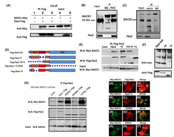
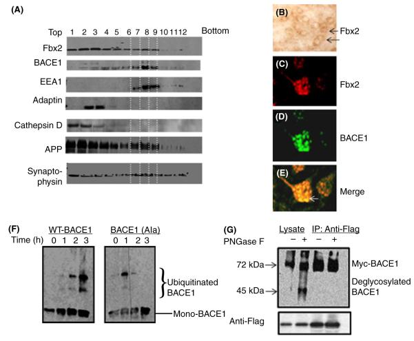
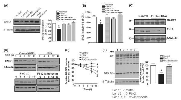
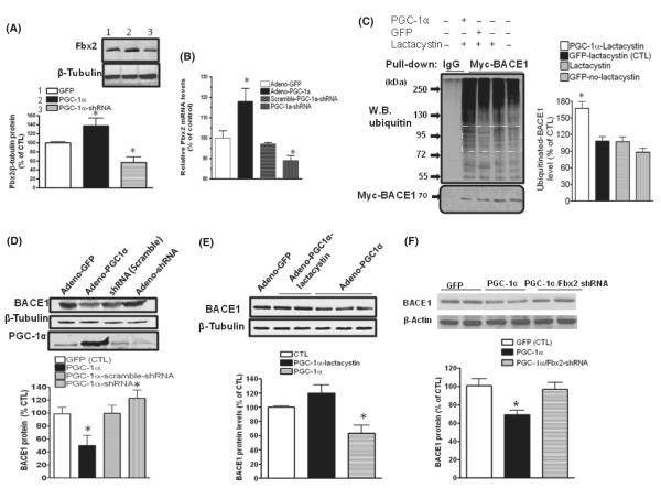
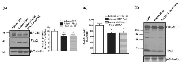
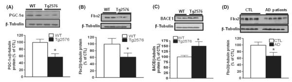
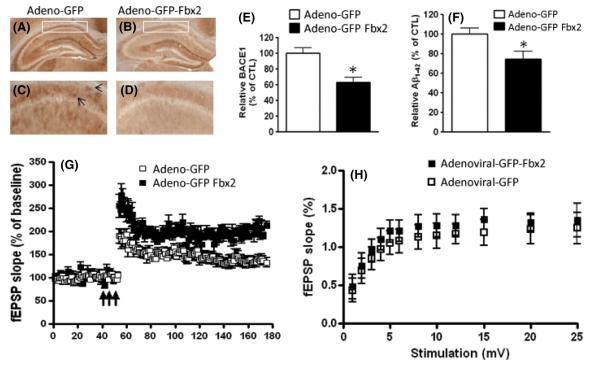
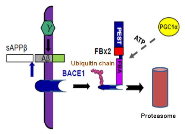
BACE1 (β-secretase) plays a central role in the β-amyloidogenesis of Alzheimer's disease (AD). The ubiquitin-proteasome system, a major intracellular protein quality control system, has been implicated recently in BACE1 metabolism. We report that the SCF(Fbx2) -E3 ligase is involved in the binding and ubiquitination of BACE1 via its Trp 280 residue of F-box-associated domain. Physiologically, we found that Fbx2 was expressed in various intracellular organelles in brain neurons and that BACE1 is colocalized with Fbx2 and the amyloid precursor protein (APP), mainly at the early endosome and endoplasmic reticulum. The former are believed to be the major intracellular compartments where the APP is cleaved by BACE1 and β-amyloid is produced. Importantly, we found that overexpression of Fbx2 in the primary cortical and hippocampal neurons derived from Tg2576 transgenic mice significantly promoted BACE1 degradation and reduced β-amyloid production. In the search for specific endogenous modulators of Fbx2 expression, we found that PPARγ coactivator-1α (PGC-1α) was capable of promoting the degradation of BACE1 through a mechanism involving Fbx2 gene expression. Interestingly, we found that the expression of both Fbx2 and PGC-1α was significantly decreased in the brains of aging Tg2576 mice. Our in vivo studies using a mouse model of AD revealed that exogenous adenoviral Fbx2 expression in the brain significantly decreased BACE1 protein levels and activity, coincidentally reducing β-amyloid levels and rescuing synaptic deficits. Our study is the first to suggest that promoting Fbx2 in the brain may represent a novel strategy for the treatment of AD.


Figures

Fig. 1
Fig. 1
Interaction of Fbx2 with BACE1. (A) HEK293 cells stably expressing Myc-BACE1 were cotransfected with FLAG-Fbx2 plasmids. After immunoprecipitation of Fbx2–FLAG from whole-cell extracts, bound BACE1 was analyzed using an antibody against Myc. Tissue lysates from mouse (B) and human (C) brains were subjected to coimmunoprecipitation with anti-Fbx2 antibody and detected with anti-BACE1 antibody. In both tissue co-immunoprecipitations, anti-mouse or anti-rabbit IgG antibody were used as negative controls. (D) Depiction of Fbx2 and mutants. (E, F) Fbx2 binds to BACE1 through FBA (ΔC) domain. Two hundred and ninety-three cells were transiently transfected with FLAG-tagged Fbx2 variants. Cell lysates were immunoprecipitated with goat anti-FLAG antibody and the bound BACE1 was analyzed using an antibody against Myc. (G) FBA domain point mutations, Trp280, Tyr279, and K281 were transfected into 293 cells. Immunoprecipitation shows that only W280 is required for the binding. (H-T) Colocalization of Fbx2 and BACE1. 293 stable cells co-expressing Fbx2 and its variants were immunostained with anti-Myc and –FLAG antibodies and examined via confocal microscopy. White arrows indicate colocalization of Fbx2 and BACE1 (panels J and T). FBA domain deletion (panel K-M) and the W280A mutation were not colocalized with BACE1 (panels N-P) Scale bar: 10 μm. FBA, F-box-associated.
Fig. 2
Fig. 2
Subcellular localizations of the Fbx2 and BACE1 complex in brain neurons. (A) A stepwise sucrose gradient ultracentrifugation for the subcellular fraction of brain tissue. Nine hundred and fifty microliter fractions were collected and analyzed by Western blot. Fbx2 and APP are broadly expressed in lysosomes (cathepsin D) and early and late endosomes (EEA1 and adaptin) and are colocalized with BACE1, mainly in the early endosome (EEA1, lanes 7, 8, 9) and much less in the late endosome (lanes 3, 4) and lysosome (lane 1, 2). (B) Sections of brain slices immunostained by the ABC procedure with Fbx2 antibody. (C–E) Immunofluorescence staining of BACE1 and Fbx2, showing the colocalization of BACE1 and Fbx2 in brain slices (white arrow). (F) In vitro ubiquitination of BACE1 by the SCFFbx2 E3-ligase system. Fbx2 enhances WT BACE1 ubiquitination (left pane) but not mutant BACE1 ubiquitination (right panel). (G) N-glycosylation of BACE1 is required for the Fbx2-mediated binding. Cell lysate from cells treated (+) with PNGase F at 37 °C for 1 h or from controls (−) were immunoprecipitated with anti-FLAG antibody and probed with anti-Myc antibody. Only glycosylated BACE1 can be immunoprecipitated by Fbx2. ABC, avidin–biotin–peroxidase complex; APP, amyloid precursor protein. WT, wild-type.
Fig. 3
Fig. 3
Fbx2 promotes BACE1 protein degradation. (A) HEK293 cells stably expressing human BACE1 protein were cotransfected with Fbx2 and treated with lactacystin. The levels of BACE1 protein were probed with anti-BACE1 antibody. Equal sample loading was normalized with antibody to β-tubulin (right panel). Protein amounts were quantified by Image J and represented in the graph. Data are means ± SEM (error bar) of the results from three independent experiments. One-way anova reveals the differences between each condition with control group; *P < 0.05. (B) BACE1 enzyme activity from the same cell lysate was measured using a BACE1-FRET kit (EMD biology). Data are means ± SEM, one-way anova,*P < 0.05 n = 5. (C) Hippocampal neurons derived from Tg2576 embryos infected with adenoviral GFP or retroviral Fbx2 shRNA. BACE1 protein levels were increased by silencing endogenous Fbx2. Data represent means ± SEM; n = 5 per culture; *P < 0.05 relative to adeno-GFP-infected cell cultures. (D) BACE1 stability was measured by cycloheximide (CHX) chase followed by Western blot. After 48 h of transfection, cells were treated with CHX (50 μm) and chased at the indicated time points. (E) The quantified relative levels of BACE1 against levels of β-tubulin and plotted as a percentage of the level of BACE1 at chase time t = 0. Two-way anova reveals the differences between Fbx2 and control group; *P < 0.05 compared with control group. (F) Stable HEK293 cells were transiently transfected with human APPswe and Fbx2, and treated with lactacystin. The β-secretase-cleaved APP C-terminal fragments CTF99 were analyzed by Western blot using anti-APP C-terminal antibody (APP 8344). Fbx2 reduces C99 cleavage (lanes 3, 4, 5). APP, amyloid precursor protein; FRET, fluorescence resonance energy transfer.
Fig. 4
Fig. 4
PGC-1α-mediated BACE1 degradation through Fbx2. Hippocampal neurons derived from Tg2576 embryos were infected with adenoviral GFP, adenoviral PGC-1α, PGC-1α shRNA or scrambled shRNA, respectively. (A) Forty-eight hours after infection, Fbx2 protein levels were probed using anti-Fbx2 antibody. PGC-1α markedly increases Fbx2 protein expression (lane 2), while adenoviral-PGC-1α shRNA reduces the Fbx2 levels (lane 3) (N = 5, means ± SEM, *P < 0.05. (B) Total RNA was extracted from transfected cells. Levels of Fbx2 mRNA were quantified by qRT-PCR (n = 5 independent studies; *P < 0.05). (C) PGC-1α promotes BACE1 ubiquitination. HEK293 cells stably expressing Myc-BACE1 were infected with adenoviral-PGC-1α or adenoviral-GFP constructs and treated with 5 μm lactacystin. BACE1 was immunoprecipitated with an antibody against Myc, The ubiquitinated BACE1 was probed with anti-ubiquitin antibody. The levels of ubiquitinated BACE1 levels in the square area were quantified and represented in the graph (right panel). Data are means ± SEM of the results from two independent experiments; *P < 0.05 compared with adenoviral-GFP/lactacystin-treated cells (control). (D) Silencing endogenous PGC-1α blocks BACE1 degradation. Neurons were infected with adenoviral GFP, adenoviral PGC-1α, and adenoviral PGC-1α shRNA; (E) lactacystin blocked the effects of PGC-1α on BACE1 degradation. (F) Silencing Fbx2 diminishes the effects of PGC-1α on BACE1 degradation. (n = 5, data are means ± SEM; n = 5 per culture; *P < 0.05 relative to adenoviral-GFP-infected cell cultures).
Fig. 5
Fig. 5
Fbx2 acts on the downstream of PGC-1α. (A) Silencing endogenous PGC-1α did not affect the role of Fbx2 in the BACE1 degradation in primary cultured neurons from E16 Tg2576 embryos. BACE1 protein was probed by Western blot with anti-BACE1 antibody and normalized by β-tubulin (left panel). The right panel shows quantification of BACE1 protein levels. Data are means ± SEM; n = 5, *P < 0.05 compared with control. (B) BACE1 activity was measured in (A) and quantified as a percent of control; *P < 0.05. (C) Full APP and BACE1 cleavage product APP-C terminal fragment C99 were analyzed by immunoblots with 6E10 antibody. APP, amyloid precursor protein.
Fig. 6
Fig. 6
Fbx2 and PGC-1α expression are compromised in Tg2576 mice and human AD brains. (A) PGC-1α protein levels are reduced in the hippocampal formation of 12- to 14-month-old Tg2576 mice compared to age- and gender-matched WT littermates. (B and D) Fbx2 protein levels are decreased in 12-month-old Tg2576 mice and in the postmortem brains of patients with AD (parahippocampal gyrus; BA 36), respectively. (C) BACE1 protein levels are elevated in the hippocampal formation of 12- to 14-month-old Tg2576 mice compared to age- and gender-matched WT littermates. The lower panels in D, E, F, and G showed the quantification of immunoreactivity. Student’s t-test was used for statistical analysis. *P < 0.05, n = 5 per group. AD, Alzheimer’s disease; WT, wild-type.
Fig. 7
Fig. 7
Stereotaxic injection of Fbx2 reduces BACE1 levels and improves synaptic function. Mouse brain specimens were analyzed 4 weeks after injection of either adenoviral GFP-Fbx2 or control adenoviral-GFP constructs. (A, B) Representative micrographs of the BACE1 immunoreactive signal in the CA1 pyramidal layer of the hippocampal formation of Tg2576 mice 4 weeks after injection. Boxed areas of the CA1 region shown in (A) and (B) are enlarged in (C) and (D), respectively. Arrows show BACE1 immunostaining signal. (E) Quantitative assessment of the BACE1 immunoreactive signal in (C) and (D) (n = 15 slices/group, three consecutive sections/mouse, five mice in each group; *P < 0.05). (F) ELISA determination of Aβ1-42 levels in the hippocampal formation of Tg2576 mice 4 weeks after injection. Values are expressed as means ± SEM, n = 5 per group; *P < 0.05, Student t-test relative to control adenoviral GFP injection. (G) Analysis of long-term potentiation (LTP) in the hippocampal Schaffer collateral/CA1 region of Tg2576 mice shows that Fbx2 significantly increased LTP compared with adenoviral-GFP-treated Tg mice (these experiments were interleaved with those of adenoviral-GFP-treated mice). Arrows indicate time and pattern of the tetani. All data shown are means ± SEM; n = 10 slices from five mice in each group; two-way anova following post hoc analysis reveals P < 0.01. (H) Basal synaptic transmission at the connection of hippocampal slices from 12- to 14-month-old Tg2576 mice was not affected by Fbx2 4 weeks after injection (n = 10 slices from five mice in each group, two-way anova; P > 0.05).
Fig. 8
Fig. 8
Working hypothesis of Fbx2 facilitating BACE1 degradation. In the AD brain, BACE1 is essential to cleave APP to generate Aβ. Fbx2 binds BACE1 through the F-box domain and promotes BACE1 ubiquitination and proteasomal degradation. PGC-1α mediated BACE1 metabolism through the enhancement of Fbx2 gene/protein expression and further reduced Aβ production. AD, Alzheimer’s disease; APP, amyloid precursor protein.

Similar articles

Cited by

References

    1. Anderson RM, Barger JL, Edwards MG, Braun KH, O’Connor CE, Prolla TA, Weindruch R. Dynamic regulation of PGC-1αlpha localization and turnover implicates mitochondrial adaptation in longevity and the stress response. Aging Cell. 2008;7:10. - PMC - PubMed
    1. Cardozo T, Pagano M. The SCF ubiquitin ligase: insights into a molecular machine. Nat. Rev. Mol. Cell Biol. 2004;5:739–751. - PubMed
    1. Chapman PF, White GL, Jones MW, Cooper-Blacketer D, Marshall VJ, Irizarry M, Younkin L, Good MA, Bliss TV, Hyman BT, Younkin SG, Hsiao KK. Impaired synaptic plasticity and learning in aged amyloid precursor protein transgenic mice. Nat. Neurosci. 1999;2:271–276. - PubMed
    1. Ciechanover A, Brundin P. The ubiquitin proteasome system in neurodegenerative diseases: sometimes the chicken, sometimes the egg. Neuron. 2003;40:427–446. - PubMed
    1. Cole S, Vassar R. The Alzheimer’s disease beta-secretase enzyme, BACE1. Mol. Neurodegener. 2007;2:22. - PMC - PubMed

Publication types

MeSH terms