Skin wound healing in diabetic β6 integrin-deficient mice
- PMID: 20854469
- PMCID: PMC2964129
- DOI: 10.1111/j.1600-0463.2010.02654.x
Skin wound healing in diabetic β6 integrin-deficient mice
Abstract
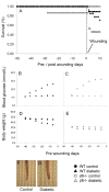
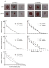
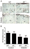
Integrin αvβ6 is a heterodimeric cell surface receptor, which is absent from the normal epithelium, but is expressed in wound-edge keratinocytes during re-epithelialization. However, the function of the αvβ6 integrin in wound repair remains unclear. Impaired wound healing in patients with diabetes constitutes a major clinical problem worldwide and has been associated with the accumulation of advanced glycated endproducts (AGEs) in the tissues. AGEs may account for aberrant interactions between integrin receptors and their extracellular matrix ligands such as fibronectin (FN). In this study, we compared healing of experimental excisional skin wounds in wild-type (WT) and β6-knockout (β6(-/-) ) mice with streptozotocin-induced diabetes. Results showed that diabetic β6(-/-) mice had a significant delay in early wound closure rate compared with diabetic WT mice, suggesting that αvβ6 integrin may serve as a protective role in re-epithelialization of diabetic wounds. To mimic the glycosylated wound matrix, we generated a methylglyoxal (MG)-glycated variant of FN. Keratinocytes utilized αvβ6 and β1 integrins for spreading on both non-glycated and FN-MG, but their spreading was reduced on FN-MG. These findings indicated that glycation of FN and possibly other integrin ligands could hamper keratinocyte interactions with the provisional matrix proteins during re-epithelialization of diabetic wounds.
© 2010 The Authors. Journal Compilation © 2010 APMIS.
Conflict of interest statement
The authors declare no conflict of interest.
Figures


Similar articles
-
Mice lacking beta6 integrin in skin show accelerated wound repair in dexamethasone impaired wound healing model.Wound Repair Regen. 2009 May-Jun;17(3):326-39. doi: 10.1111/j.1524-475X.2009.00480.x. Wound Repair Regen. 2009. PMID: 19660040
-
The alphavbeta6 integrin plays a role in compromised epidermal wound healing.Wound Repair Regen. 2006 May-Jun;14(3):289-97. doi: 10.1111/j.1743-6109.2006.00123.x. Wound Repair Regen. 2006. PMID: 16808807
-
Increased expression of beta6-integrin in skin leads to spontaneous development of chronic wounds.Am J Pathol. 2004 Jan;164(1):229-42. doi: 10.1016/s0002-9440(10)63113-6. Am J Pathol. 2004. PMID: 14695336 Free PMC article.
-
Fibronectin matrix deposition and fibronectin receptor expression in healing and normal skin.J Invest Dermatol. 1990 Jun;94(6 Suppl):128S-134S. doi: 10.1111/1523-1747.ep12876104. J Invest Dermatol. 1990. PMID: 2161886 Review.
-
Activation of keratinocyte fibronectin receptor function during cutaneous wound healing.J Cell Sci Suppl. 1987;8:199-209. doi: 10.1242/jcs.1987.supplement_8.11. J Cell Sci Suppl. 1987. PMID: 2460476 Review.
Cited by
-
FOXO1 differentially regulates both normal and diabetic wound healing.J Cell Biol. 2015 Apr 27;209(2):289-303. doi: 10.1083/jcb.201409032. J Cell Biol. 2015. PMID: 25918228 Free PMC article.
-
Mulberry-Derived 1-Deoxynojirimycin Prevents Type 2 Diabetes Mellitus Progression via Modulation of Retinol-Binding Protein 4 and Haptoglobin.Nutrients. 2022 Oct 28;14(21):4538. doi: 10.3390/nu14214538. Nutrients. 2022. PMID: 36364802 Free PMC article.
-
Cellular and molecular mechanisms of skin wound healing.Nat Rev Mol Cell Biol. 2024 Aug;25(8):599-616. doi: 10.1038/s41580-024-00715-1. Epub 2024 Mar 25. Nat Rev Mol Cell Biol. 2024. PMID: 38528155 Review.
-
Healing Ability of Central Corneal Epithelium in Rabbit Ocular Surface Injury Models.Transl Vis Sci Technol. 2022 Jun 1;11(6):28. doi: 10.1167/tvst.11.6.28. Transl Vis Sci Technol. 2022. PMID: 35771535 Free PMC article.
-
Circulating Hypoxia Responsive microRNAs (HRMs) and Wound Healing Potentials of Green Tea in Diabetic and Nondiabetic Rat Models.Evid Based Complement Alternat Med. 2019 Jan 1;2019:9019253. doi: 10.1155/2019/9019253. eCollection 2019. Evid Based Complement Alternat Med. 2019. PMID: 30713578 Free PMC article.
References
-
- Wild S, Roglic G, Green A, Sicree R, King H. Global prevalence of diabetes: estimates for the year 2000 and projections for 2030. Diabetes Care. 2004;27:1047–53. - PubMed
-
- Galkowska H, Wojewodzka U, Olszewski WL. Chemokines, cytokines, and growth factors in keratinocytes and dermal endothelial cells in the margin of chronic diabetic foot ulcers. Wound Repair Regen. 2006;14:558–65. - PubMed
-
- Falanga V. Wound healing and its impairment in the diabetic foot. Lancet. 2005;366:1736–43. - PubMed
-
- Galiano RD, Tepper OM, Pelo CR, Bhatt KA, Callaghan M, Bastidas N, Bunting S, Steinmetz HG, Gurtner GC. Topical vascular endothelial growth factor accelerates diabetic wound healing through increased angiogenesis and by mobilizing and recruiting bone marrow-derived cells. Am J Pathol. 2004;164:1935–47. - PMC - PubMed
Publication types
MeSH terms
Substances
Grants and funding
LinkOut - more resources
Full Text Sources
Other Literature Sources
Miscellaneous

