. 2010 Sep 20:11:35.
doi: 10.1186/1471-2091-11-35.

Molecular characterization of tlyA gene product, Rv1694 of Mycobacterium tuberculosis: a non-conventional hemolysin and a ribosomal RNA methyl transferase

Affiliations

Molecular characterization of tlyA gene product, Rv1694 of Mycobacterium tuberculosis: a non-conventional hemolysin and a ribosomal RNA methyl transferase

Aejazur Rahman et al. BMC Biochem. .

Abstract

Background: Mycobacterium tuberculosis is a virulent bacillus causing tuberculosis, a disease responsible for million deaths each year worldwide. In order to understand its mechanism of pathogenesis in humans and to help control tuberculosis, functions of numerous Mycobacterium tuberculosis genes are being characterized. In this study we report the dual functionality of tlyA gene product of Mycobacterium tuberculosis annotated as Rv1694, a 268 amino acid long basic protein.

Results: The recombinant purified Rv1694 protein was found to exhibit hemolytic activity in vitro. It showed concentration and time-dependent hemolysis of rabbit and human erythrocytes. Multiple oligomeric forms (dimers to heptamers) of this protein were seen on the membranes of the lysed erythrocytes. Like the oligomers of conventional, well-known, pore-forming toxins, the oligomers of Rv1694 were found to be resistant to heat and SDS, but were susceptible to reducing agents like β-mercaptoethanol as it had abolished the hemolytic activity of Rv1694 indicating the role of disulfide bond(s). The Rv1694 generated de novo by in vitro transcription and translation also exhibited unambiguous hemolysis confirming the self assembly and oligomerization properties of this protein. Limited proteolytic digestion of this protein has revealed that the amino terminus is susceptible while in solution but is protected in presence of membrane. Striking feature of Rv1694 is its presence on the cell wall of E. coli as visualized by confocal microscopy. The surface expression is consistent with the contact dependent haemolytic ability of E. coli expressing this protein. Also, immune serum specific to this protein inhibits the contact dependent hemolysis. Moreover, Rv1694 protein binds to and forms stable oligomers on the macrophage phagosomal membranes. In addition to all these properties, E. coli expressing Rv1694 was found to be susceptible to the antibiotic capreomycin as its growth was significantly slower than mock vector transformed E. coli. The S30 extract of E. coli expressing the Rv1694 had poor translational activity in presence of capreomycin, further confirming its methylation activity. Finally, incorporation of methyl group of [3H]-S-adenosylmethionine in isolated ribosomes also confirmed its methylation activity.

Conclusions: The Rv1694 has an unusual dual activity. It appears to contain two diverse functions such as haemolytic activity and ribosomal RNA methylation activity. It is possible that the haemolytic activity might be relevant to intra-cellular compartments such as phagosomes rather than cell lysis of erythrocytes and the self-assembly trait may have a potential role after successful entry into macrophages by Mycobacterium tuberculosis.


Figures

Figure 1
Figure 1
(A) Multiple sequence alignment of Rv1694 with pore forming hemolysins. Multiple sequence alignment (BCM search launcher) of Rv1694 (TlyA) of M. tb with TlyA sequences of S. hyodysenteria and H. pylori. TlyA of Mtb shows 58% homology with TlyA of both S. hyodysenteria (38% identity) and H. pylori (32% identity). TlyAs of all these bacteria also have K-D-K-E motif (indicated with inverted arrow) (▼) which were predicted to be signature motiffs of ribosomal RNA methyltransferases. (B) Alignment with ribosomal RNA methyltransferase: Homology of Rv1694 with ribosomal RNA methyltransferase (FtsJ/RrmJ) from E. coli (Multiple sequence alignment with ClustalW). TlyA of Mtb shows 48% homology with FtsJ/RrmJ.
Figure 2
Figure 2
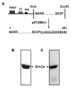
(A) Cloning of Rv1694. Cloning strategy of Rv1694 gene in E. coli expression vectors, pT7Nc and pET28a(+). The latter vector provides a C-terminal 6-histidine tag. (B) Purification of recombinant Rv1694: Expression of 6-histidine-Rv1694 in BL21(DE3) CodonPlus-RIPL E. coli after induction with 1 mM IPTG which was purified on Ni-NTA affinity column. Protein sample was electrophoresed on 12% SDS-PAGE and stained with coomassie blue R-250. The purified Rv1694 routinely exhibited >95% purity. (C) Immuno detection of Rv1694: Purified Rv1694 was electrophoresed on 12% SDS-PAGE and the immuno-probed with Rv1694 specific immune rabbit serum (at 1:1000 dilution) and the secondary antibody was anti-rabbit IgG HRP- antibody (1:2000 dilution). The specific protein band of Rv1694 can be seen at 30 kDa.
Figure 3
Figure 3
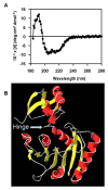
(A) Circular-Dichroic spectra of Rv1694. Rv1694-6-histidine tag fusion protein (0.5 mg/ml in 10 mM Na-Phosphate buffer, pH 7.4) was used for obtaining CD spectra. CD data, as interpreted from Yang's calculation, shows that Rv1694 has 23% α-helix, 32.8% β-sheet, 7.7% turns and 36.5% random coil. (B) Model of Rv1694: Model of Rv1694 was built using the putative hemolysin from Streptococcus thermophilus, using 3HP7.pdb as template with the help of Swiss Model Server [33]. The Rv1694 has a typical fold of a ribosomal RNA polymerase consisting of 7 β-sheets surrounded by 5 α-helices. Please note that the amino acids 246-268(QTD...EGP) are not present in the depicted model. The white arrow points the region containing basic amino acids between the domains susceptible to proteases like trypsin/Proteinase K.
Figure 4
Figure 4
(A) Contact dependent hemolysis of Rv1694. Rv1694 (with and w/o His-tag) and mock vector transformed E. coli were examined for contact dependent lysis of rabbit RBC. E. coli (~107) cells and rabbit erythrocytes (~105) were mixed and briefly centrifuged to ensure close contact between bacteria and RBC. The resultant mixture of cells was incubated at 37°C for 24 to 30 hours. Degree of lysis was monitored by measuring the absorbance at 540 nm of a cell-free supernatant. UI indicates uninduced E. coli and groups labeled with 1-5 were harvested time in hour(s) from induction (with 1.0 mM IPTG) time point. Error bars represent standard deviation of two independent experiments. (B) Hemolytic activity of the purified Rv1694: Specific hemolytic activity of purified Rv1694 (40 μg/ml) was carried out by two-fold serial dilution of Rv1694 which was mixed with rabbit RBC (1.5%). After 24 hrs of incubation, the absorbance was measured at 540 nm for release of hemoglobin. At ~50% hemolysis, the protein concentration was 18.0 μmug/ml. In presence of thiol-reducing agent, the hemolytic activity was 3.6 fold lesser than the maximum activity of Rv1694. Total haemoglobin release was obtained by lysing the RBC with deionized water. Error bars represent standard deviation from three independent experiments. (C) Hemolytic activity of Rv1694 generated by coupled in vitro transcription and translation: Rv1694 generated by coupled in vitro transcription and translation (~5.0 out of 50.0 μl reaction mix) was mixed with 100 μl of 0.3% rRBC in a 96 well plate as described earlier and the absorbance at 590 nm for rRBC lysis was noted every 10 minutes. As a positive control, we have also translated staphylococcal α-HL and followed its hemolysis.
Figure 5
Figure 5
(A) Immuno-fluorescence localization of Rv1694 on E. coli: E. coli transformed with Rv1694 or pET28a+ was immunostained with immune rabbit serum followed by detection with Cy2 conjugated goat anti-rabbit IgG (green fluorescent). Left side of the panels show the Rv1694 localization (Green), middle panels show the DAPI stained bacteria (Blue) and right panels show the merged images. (B) Inhibition of contact dependent hemolysis by Immune rabbit serum: Rv1694 transformed E. coli was incubated with and without pre-immune/immune rabbit serum as described for Figure 4A. Error bars represent standard deviation of two independent experiments.
Figure 6
Figure 6
(A) Binding and oligomerization of Rv1694 on rRBC membrane. Various amounts of purified Rv1694 were incubated with 2% rRBC for 30 min and the resultant blot was immuno probed with anti-6-histidine-antibody. Lanes indicated with RBC, Sup and numbers respectively indicate rRBC membrane, purified Rv1694, supernatant of rRBC, rRBC incubated with 5.0 μg, 10.0 μg, 20.0 μg and 30.0 μg Rv1694 respectively. (B) Oligomerization of Rv1694 on RBC membrane in the absence of reducing agents: Oligomerization of Rv1694 (10 μmug) on rRBC (2%) was carried out as described in methods section. The samples were washed and dissolved in 4% SDS and 5× laemmli sample buffer without β-mercaptoethanol and immuno probed with anti-6-histidine-antibody. Lane 1. Rv1694 purified protein; Lanes 2-5 show the oligomers of Rv1694 on the RBC membrane at indicated temperatures (°C). (C) Binding and oligomerization of Rv1694 protein on the Phagosomal membrane: Aliquots were taken out at indicated time points from the Rv1694 incubated phagosomal preparations, electrophoresed on 8% SDS-PAGE (non-reducing), immuno probed with anti-6-histidine-antibody. Lane 1: Phagosomal preparation alone; Lane 2: Unboiled Rv1694; Lanes 3 to 5: Unboiled samples of phagosomal preparations and Rv1694 incubated for 10.0, 30.0, and 60.0 min respectively. Lane 6: Boiled Rv1694 protein; Lane 7: Boiled sample of lane 5. Labels viz., M, O3, O5, O6 and O7 indicate monomers, trimers, pentamers, hexamers and heptamers respectively. The blot was stripped and re-probed with anti-LAMP-1 antibody to ascertain the phagosomal preparation. The data shown is one of the three independent experiments.
Figure 7
Figure 7
(A) & (B) Limited proteolysis of Rv1694 with Proteinase K. Rv1694 was partially digested with Proteinase K for indicated times, electrophoresed on 12% SDS-PAGE, stained with coomassie brilliant blue R-250. The lanes were marked to indicate M for protein molecular weight markers, numbers represent the time points: 0 for Rv1694 protein only and lanes marked with 0.5, 1, 2, 5, 10, 20 and 30 respectively represent Proteinase K treatment times in minutes. The arrow on the right indicates the stable domains of Rv1694. The panel (B) was obtained by probing with anti-6-histidine monoclonal antibody. (C) Limited proteolysis of Rv1694 bound to rRBC membranes: Purified Rv1694 protein and rRBC were incubated for 30 min and subjected to digestion with Proteinase K for indicated periods of time. At the end of the incubation, the rRBC were mixed with 2 mM PMSF, washed twice with hypotonic buffer, solublized the RBC membranes and were electrophoresed on 12% SDS-PAGE, probed with anti-6-histidine-monoclonal antibody. The lanes marked with Rv1694 and RBC respectively represent Rv1694 protein only and rRBC membrane only. Lanes indicated with 0.5, 1, 2, 5, 10, 20 and 30 respectively represents the Proteinase K treatment times. Results shown are one of the two independent experiments.
Figure 8
Figure 8
(A) Growth of E. coli expressing Rv1694 in presence of capreomycin: Growth of E. coli expressing Rv1694 in the absence (●) and presence (▼) of capreomycin (100 μmug/ml), and pET28a+ transformed E. coli (○). Error bars represent standard errors from two independent experiments. (B) Inhibition of coupled in vitro transcription-translation by capreomycin: S30 extracts of Rv1694 or pT7Nc transformed E. Coli BL21(DE3) were prepared and in vitro transcription and translation reactions were carried out with either 16 or 80 ng/ml capreomycin. The hemolytic activity of staphylococcal α-hemolysin (used as a reporter) was monitored at OD595. All points for all curves were calculated with respect to 100% lysis of rRBCs and average of three independent experiments is shown. Various symbols represent (●) Rv1694 without capreomycin; (○) mock vector without capreomycin; (▼) Rv1694 at 16 ng/ml capreomycin; (∆) mock vector at 16 ng/ml capreomycin; (■) Rv1694 80 at ng/ml capreomycin; (□) mock vector at 80 ng/ml capreomycin. (C) In vitro methylation activity of purified Rv1694 protein on isolated Ribosome: Methylation of E. coli ribosomes in presence or absence of purified Rv1694 and [3H]S-adenosylmethionine. From left, Crossed pattern bar represents the reaction mix with native ribosome ('Nat Rib') and Rv1694 ('Prot'), horizontal pattern bar represents reaction mix with native ribosome only, open bar represents reaction mix with heat (65°C) denatured ribosome ('Den Rib') and Rv1694 and filled bar represents reaction mix with denatured ribosome only. Error bars represent standard errors from triplicate experiments.

References

    1. Cole ST, Brosch R, Parkhill J, Garnier T, Churcher C, Harris D, Gordon SV, Eiglmeier K, Gas S, Barry CE III. et al.Deciphering the biology of Mycobacterium tuberculosis from the complete genome sequence. Nature. 1998;393:537–544. doi: 10.1038/31159. - DOI - PubMed
    1. Camus JC, Pryor MJ, Medigue C, Cole ST. Re-annotation of the genome sequence of Mycobacterium tuberculosis H37Rv. Microbiology. 2002;148:2967–2973. - PubMed
    1. Betts JC, Dodson P, Quan S, Lewis AP, Thomas PJ, Duncan K, McAdam RA. Comparison of the proteome of Mycobacterium tuberculosis strain H37Rv with clinical isolate CDC 1551. Microbiology. 2000;146(Pt 12):3205–3216. - PubMed
    1. Mollenkopf HJ, Jungblut PR, Raupach B, Mattow J, Lamer S, Zimny-Arndt U, Schaible UE, Kaufmann SH. A dynamic two-dimensional polyacrylamide gel electrophoresis database: the mycobacterial proteome via Internet. Electrophoresis. 1999;20:2172–2180. doi: 10.1002/(SICI)1522-2683(19990801)20:11<2172::AID-ELPS2172>3.0.CO;2-M. - DOI - PubMed
    1. Jungblut PR, Schaible UE, Mollenkopf HJ, Zimny-Arndt U, Raupach B, Mattow J, Halada P, Lamer S, Hagens K, Kaufmann SH. Comparative proteome analysis of Mycobacterium tuberculosis and Mycobacterium bovis BCG strains: towards functional genomics of microbial pathogens. Mol Microbiol. 1999;33:1103–1117. doi: 10.1046/j.1365-2958.1999.01549.x. - DOI - PubMed

Publication types

MeSH terms

LinkOut - more resources