

Review
doi: 10.1016/j.abb.2010.09.012.
Epub 2010 Sep 18.
Structure and mechanism of enzymes involved in biosynthesis and breakdown of the phosphonates fosfomycin, dehydrophos, and phosphinothricin
Affiliations
- PMID: 20854789
- PMCID: PMC3040005
- DOI: 10.1016/j.abb.2010.09.012
Item in Clipboard
Review
Structure and mechanism of enzymes involved in biosynthesis and breakdown of the phosphonates fosfomycin, dehydrophos, and phosphinothricin
Arch Biochem Biophys.
.
Abstract
Recent years have seen a rapid increase in the mechanistic and structural information on enzymes that are involved in the biosynthesis and breakdown of naturally occurring phosphonates. This review focuses on these recent developments with an emphasis on those enzymes that have been characterized crystallographically in the past five years, including proteins involved in the biosynthesis of phosphinothricin, fosfomycin, and dehydrophos and proteins involved in resistance mechanisms.
Copyright © 2010 Elsevier Inc. All rights reserved.
Figures
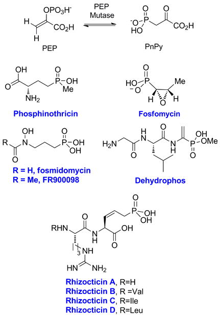
Phosphorus-carbon bond formation by PEP mutase and structures of the phosphonate natural products for which the gene clusters have been determined.
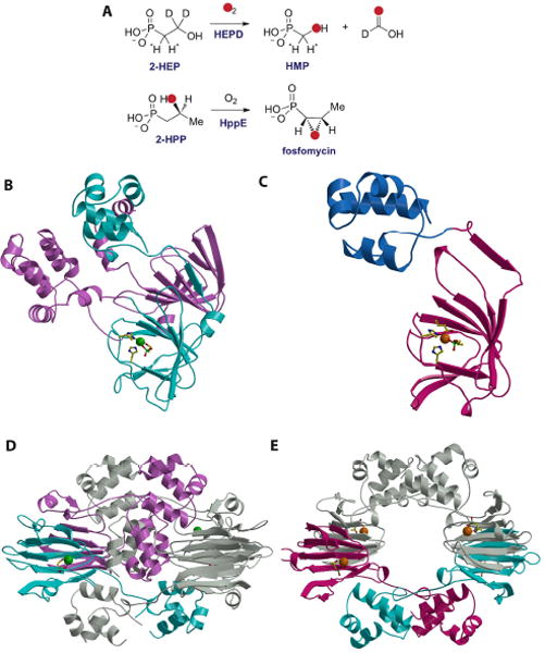
(A) Reactions catalyzed by HEPD and HppE. (B) Structure of the HEPD-HEP complex with the active metal-containing domain colored in cyan and the vestigial domain colored in pink. The bound Cd(II) is shown as a green sphere, with protein ligands and the substrate molecule shown as stick figures. (C) Structure of the HppE-fosfomycin complex with the cupin domain colored in cyan and the novel alpha domain colored in blue. The requisite Fe(II) is colored in orange with protein ligands and the substrate molecule shown as stick figures. (D) Structure of the HEPD dimer suggesting the relevance of the vestigial repeat in forming the composite active site. (E) Structure of the HppE tetramer shown in the same orientation as that for the HEPD dimer.

(A) Two proposed mechanisms for the HEPD reaction. (B) The substrate analog 1-HEP is converted to acetylphosphate by HEPD. (C) Two proposed mechanisms for the HppE reaction. The phosphonate groups are shown as dianionic but could be monoprotonated. L = undefined ligand, probably water or hydroxide.
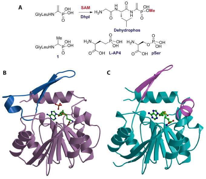
(A) Reaction catalyzed by DhpI and structures of several of the phosphonates that are also substrates for the enzyme. (B) Structure of the DhpI-SAM-sulfate complex showing the nucleotide binding Rossman fold in brown and the unusual insertions necessary for substrate binding in blue. The SAM co-factor and sulfate anion are shown as stick figures. (C) Structure of the DhpI-SAH complex with the nucleotide-binding domain colored in cyan and part of the novel insertion as well as a newly formed helix colored in pink. The helix of the insertion is disordered in the SAH complex.
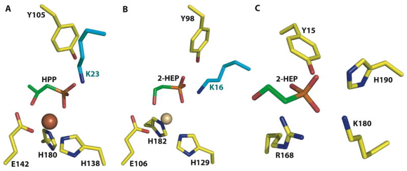
A close-up view of the actives sites of (A) HppE in complex with 2-HPP, (B) HEPD in complex with 2-HEP, and (C) DhpI in complex with 2-HEP. Despite the lack of any notable sequence similarities, both HEPD and HppE use a near identical constellation of active site residues to carry out their respective reactions. Notably, both polypeptides contain composite active sites with a catalytically essential lysine residue (colored in cyan) from a different subunit interacting with the active site. A comparison of HppE, HEPD, and DhpI co-crystal structures illustrates the chemical features that are used by functionally and structurally distinct enzymes to harbor phosphonate substrates.
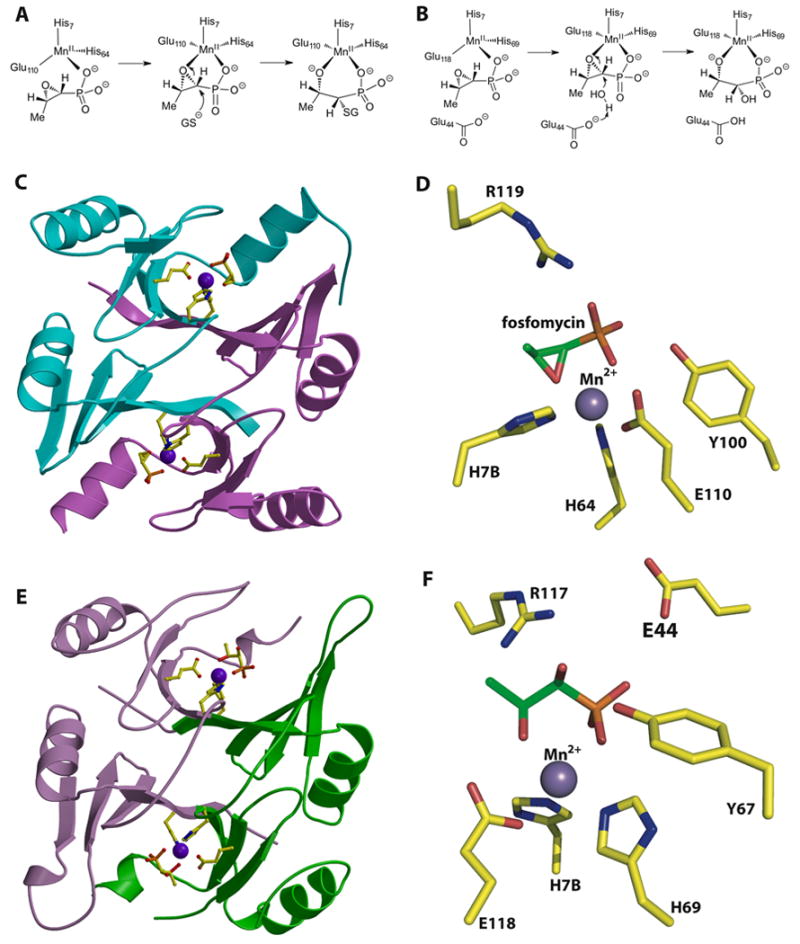
(A) Reaction catalyzed by the fosfomycin thiol transferase FosA. (B) Reaction catalyzed by the homologous FosX metalloenzyme that hydrates the antibiotic. (C) Structure of the fosfomycin inactivating enzyme FosA. (D) Close-up view of the active site of FosA. (E) Structure of the fosfomycin hydrolase FosX. (F) Close-up view of the active site of FosX revealing a glutamate residue (Glu44) that is proximal to the substrate and that is essential for catalysis. This residue is absent in FosA.
Similar articles
-
Phosphonate biosynthesis and catabolism: a treasure trove of unusual enzymology.Curr Opin Chem Biol. 2013 Aug;17(4):580-8. doi: 10.1016/j.cbpa.2013.06.018. Epub 2013 Jul 17. Curr Opin Chem Biol. 2013. PMID: 23870698 Free PMC article. Review.
-
Biosynthesis of 2-hydroxyethylphosphonate, an unexpected intermediate common to multiple phosphonate biosynthetic pathways.J Biol Chem. 2008 Aug 22;283(34):23161-8. doi: 10.1074/jbc.M801788200. Epub 2008 Jun 10. J Biol Chem. 2008. PMID: 18544530 Free PMC article.
-
Characterization and structure of DhpI, a phosphonate O-methyltransferase involved in dehydrophos biosynthesis.Proc Natl Acad Sci U S A. 2010 Oct 12;107(41):17557-62. doi: 10.1073/pnas.1006848107. Epub 2010 Sep 27. Proc Natl Acad Sci U S A. 2010. PMID: 20876132 Free PMC article.
-
Fosfomycin induced structural change in fosfomycin resistance kinases FomA: molecular dynamics and molecular docking studies.J Mol Model. 2014 May;20(5):2236. doi: 10.1007/s00894-014-2236-2. Epub 2014 Apr 27. J Mol Model. 2014. PMID: 24770549
-
The Abc of Phosphonate Breakdown: A Mechanism for Bacterial Survival.Bioessays. 2018 Nov;40(11):e1800091. doi: 10.1002/bies.201800091. Epub 2018 Sep 9. Bioessays. 2018. PMID: 30198068 Review.
Cited by
-
The Streptomyces-produced antibiotic fosfomycin is a promiscuous substrate for archaeal isopentenyl phosphate kinase.Biochemistry. 2012 Jan 31;51(4):917-25. doi: 10.1021/bi201662k. Epub 2012 Jan 11. Biochemistry. 2012. PMID: 22148590 Free PMC article.
-
GenK-catalyzed C-6' methylation in the biosynthesis of gentamicin: isolation and characterization of a cobalamin-dependent radical SAM enzyme.J Am Chem Soc. 2013 Jun 5;135(22):8093-6. doi: 10.1021/ja312641f. Epub 2013 May 21. J Am Chem Soc. 2013. PMID: 23679096 Free PMC article.
-
Structure and function of phosphonoacetaldehyde dehydrogenase: the missing link in phosphonoacetate formation.Chem Biol. 2014 Jan 16;21(1):125-35. doi: 10.1016/j.chembiol.2013.11.006. Epub 2013 Dec 19. Chem Biol. 2014. PMID: 24361046 Free PMC article.
-
Characterization of Two Late-Stage Enzymes Involved in Fosfomycin Biosynthesis in Pseudomonads.ACS Chem Biol. 2017 Feb 17;12(2):456-463. doi: 10.1021/acschembio.6b00939. Epub 2016 Dec 27. ACS Chem Biol. 2017. PMID: 27977135 Free PMC article.
-
Phosphonate biosynthesis and catabolism: a treasure trove of unusual enzymology.Curr Opin Chem Biol. 2013 Aug;17(4):580-8. doi: 10.1016/j.cbpa.2013.06.018. Epub 2013 Jul 17. Curr Opin Chem Biol. 2013. PMID: 23870698 Free PMC article. Review.
References
Publication types
MeSH terms
Substances
Grants and funding
LinkOut - more resources
Full Text Sources
Medical
Miscellaneous
