. 2010 Dec;139(6):2028-2037.e9.
doi: 10.1053/j.gastro.2010.09.005. Epub 2010 Sep 18.

Mature chief cells are cryptic progenitors for metaplasia in the stomach

Affiliations

Mature chief cells are cryptic progenitors for metaplasia in the stomach

Ki Taek Nam et al. Gastroenterology. 2010 Dec.

Abstract

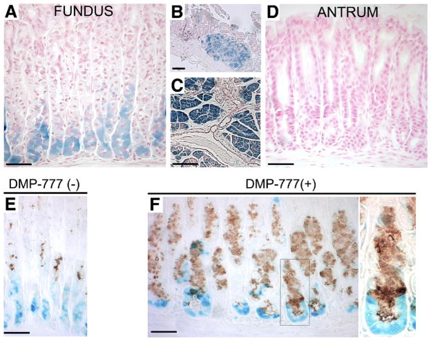
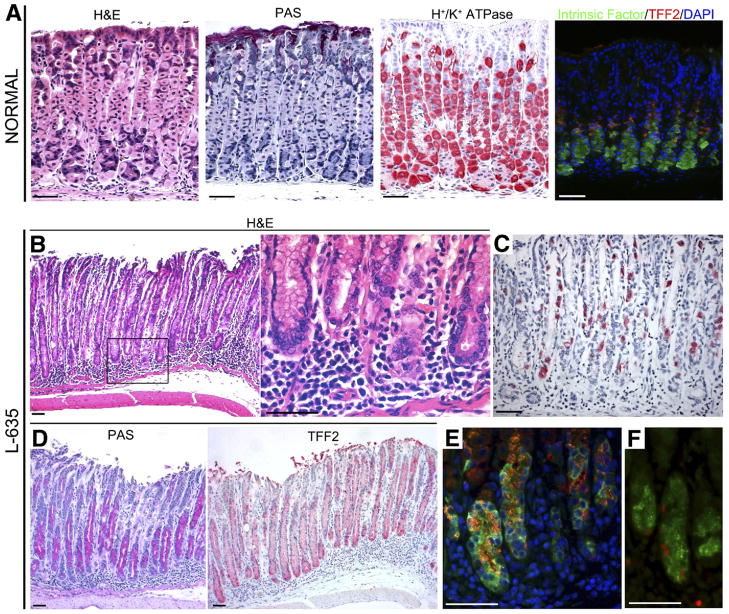
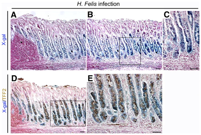
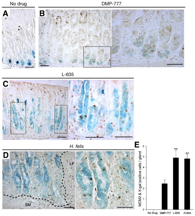
Background & aims: Gastric cancer evolves in the setting of a pathologic mucosal milieu characterized by both loss of acid-secreting parietal cells and mucous cell metaplasias. Indeed, mucous cell metaplasia is considered the critical preneoplastic lesion for gastric cancer. Previous investigations have shown that infection of mice with Helicobacter felis or induction of acute parietal cell loss with the drug DMP-777 leads to the emergence of a type of metaplasia designated spasmolytic polypeptide-expressing metaplasia (SPEM). We have hypothesized that SPEM arises from proliferating cells in gland bases, either from a cryptic progenitor cell or by transdifferentiation of mature chief cells.

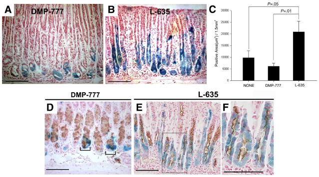
Methods: Taking advantage of the chief cell-restricted expression of Mist1-Cre-ER(T2), we used lineage mapping to examine whether SPEM lineages were derived from chief cells in 3 independent models of induction by DMP-777 treatment, L-635 treatment, or H felis infection.

Results: Treatment of mice with L-635 for 3 days led to rapid parietal cell loss, induction of a prominent inflammatory infiltrate, and emergence of SPEM. In all 3 models, SPEM developed, at least in part, from transdifferentiation of chief cells. We further found that acute parietal cell loss in the setting of inflammation (L-635 treatment) led to more rapid induction and expansion of SPEM derived from transdifferentiation of chief cells.

Conclusions: These studies provide direct evidence by lineage tracing that SPEM evolves from differentiated chief cells. Thus, mature gastric chief cells have the ability to act as cryptic progenitors and reacquire proliferative capacity within the context of mucosal injury and inflammation.


Conflict of interest statement

Conflicts of interest

The authors disclose no conflicts.

Figures

Figure 1
Figure 1
Mist1CreER/+/Rosa26RLacZ mice and DMP-777 treatment. (A–D) β-galactosidase expression in Mist1CreER/+/Rosa26RLacZ mice was induced by tamoxifen treatment. β-galactosidase activity was detected with incubation of tissue with X-gal. In mice receiving tamoxifen, strong X-gal staining (blue) was observed in chief cells of the (A) gastric fundus, (B) Brunner’s gland cells, and (C) pancreatic acinar cells, but no staining was observed in the (D) gastric antrum. (E) Stomach of a Mist1CreER/+/Rosa26RLacZ mouse treated with tamoxifen and then killed 10 days after the last tamoxifen injection stained with X-gal and antibodies against TFF2 (brown). Note the complete separation of TFF2 immunostaining mucous neck cells and X-gal–staining Mist1-expressing chief cells. (F) Tamoxifen-treated mice receiving further DMP-777 treatment. DMP-777 induced a marked loss of parietal cells and prominent SPEM, which co-stained for X-gal and TFF2. Right panel: higher magnification view. Note the co-staining of β-galactosidase–expressing, X-gal–stained cells at the bases of glands with antibodies against TFF2. Bar, 50 μm.
Figure 2
Figure 2
Characteristics of stomachs from C57BL/6 mice treated with L-635. (A) Cell lineage markers in normal mouse fundus. First panel: H&E staining; second panel: periodic acid–Schiff (PAS) staining; third panel: H+/K+-ATPase staining for parietal cells; and fourth panel: dual-immunofluorescence staining for both intrinsic factor and TFF2. A few dual-stained cells were located at the junction between the mucous neck cell and chief cell zones. Green, intrinsic factor for chief cell; red, TFF2; blue, 4,6-diamidino-2-phenylindole (DAPI). (B–F) Extensive SPEM accompanying inflammation induced by L-635. (B) H&E staining of gastric mucosa from a mouse treated with L-635 showing metaplasia with parietal cell loss and a prominent submucosal and intramucosal inflammatory infiltrate. Right panel: higher magnification view. (D) PAS staining showing prominent mucous cell metaplasia. (C) H+/K+-ATPase staining showing marked loss of parietal cells. (D) Immunostaining for TFF2 (red) in L-635–treated mice showing a marked TFF2-staining mucous cell metaplasia that dominated the fundic mucosa. (E) Dual-immunofluorescence staining for both intrinsic factor and TFF2 showing cells with dual expression of both TFF2 and intrinsic factor at the bases of fundic glands. Green, intrinsic factor; red, TFF2; blue, DAPI. (F) L-635–induced metaplastic cells also were strongly stained for Ki-67, indicating the adoption of proliferative capacity in the metaplastic cells. Green, intrinsic factor; red, Ki-67. Bar, 50 μm.
Figure 3
Figure 3
Mist1CreER/+/Rosa26RLacZ mice treated with DMP-777 or L-635. (A and B) Mist1 cell lineage analysis (X-gal staining) after treatment with either (A) DMP-777 or (B) L-635 for inducing SPEM. (C) β-galactosidase–expressing cells were quantitated as an X-gal–stained area per 1.5 mm2 of total mucosal tissue (±standard error of the mean). Compared with both untreated animals (n = 4) and DMP-777–treated mice (n = 8), L-635 treatment (n = 6) caused a significant expansion of the number of X-gal–stained cells. (D–F) Immunostaining for TFF2 in X-gal–stained sections from DMP-777 and L-635–treated mice. Brown (3,3-diaminobenzidine) = TFF2. (D) DMP-777 induced a marked loss of parietal cells and prominent SPEM, with co-staining for X-gal and TFF2 at the bases of glands (brackets). (E) TFF2 expression was observed throughout the X-gal–stained cells in mice treated with L-635. (F) Higher magnification view of panel E. Bar, 100 μm.
Figure 4
Figure 4
Lineage mapping of SPEM in 6-month-old H felis–infected Mist1CreER/+/Rosa26RLacZ mice. Sections of fundic mucosa stained with (A–C) X-gal or dual stained for (D and E) X-gal and TFF2 (brown). Chronic H felis infection caused an expansion of the number of X-gal–stained cells. (A) Note the lymphoid follicle and inflammatory cell infiltration in the mucosa. (B) Mucosa adjacent to section in panel A. (C) Higher magnification view of panel B. (D) Dual immunostaining for TFF2 in X-gal–stained sections. Brown (3,3-diaminobenzidine) = TFF2. TFF2 expression was observed throughout the X-gal–stained cells in H felis–infected mice. (E) Higher magnification view of panel D. Bar, 50 μm.
Figure 5
Figure 5
Proliferative cell lineage analysis in SPEM induced by DMP-777, L-635, and H felis infection in Mist1CreER/+/Rosa26RLacZ mice. Proliferating cells in mice were visualized with antibodies against MCM2. (A) MCM2 stained only proliferating normal progenitor cells in the upper fundic gland neck, whereas no proliferating cells were observed in the X-gal–staining chief cells (n = 4). (B) In DMP-777–treated mice (n = 8), we observed MCM2 expression in X-gal–staining cells at the bases of fundic glands. Right panel: higher magnification view. (C) In mice treated with L-635 (n = 6), MCM2 staining was observed in X-gal–staining cells along the entire gland length. Right 2 panels: higher magnification views. (D) In H felis–infected mice (n = 6), MCM2-staining nuclei were present in X-gal–staining cells throughout the length of the glands. SM, submucosa; LF, lymphoid follicle. Right panel: higher magnification view. (E) MCM2-positive proliferative cells were quantitated (±standard error of the mean). The number of proliferating chief cell-derived, X-gal–positive cells in L-635–treated animals and H felis–infected mice was significantly greater than in DMP-777–treated mice. **P < .01. Bar, 50 μm.

Similar articles

Cited by

References

    1. Karam SM, Leblond CP. Dynamics of epithelial cells in the corpus of the mouse stomach. I. Identification of proliferative cell types and pinpointing of the stem cell. Anat Rec. 1993;236:259–279. - PubMed
    1. Karam SM, Leblond CP. Dynamics of epithelial cells in the corpus of the mouse stomach. III. Inward migration of neck cells followed by progressive transformation into zymogenic cells. Anat Rec. 1993;236:297–313. - PubMed
    1. Correa P. A human model of gastric carcinogenesis. Cancer Res. 1988;48:3554–3560. - PubMed
    1. Schmidt PH, Lee JR, Joshi V, et al. Identification of a metaplastic cell lineage associated with human gastric adenocarcinoma. Lab Invest. 1999;79:639–646. - PubMed
    1. El-Zimaity HM, Ramchatesingh J, Saeed MA, et al. Gastric intestinal metaplasia: subtypes and natural history. J Clin Pathol. 2001;54:679–683. - PMC - PubMed

Publication types

MeSH terms

Substances