Neonatal gene transfer of Serca2a delays onset of hypertrophic remodeling and improves function in familial hypertrophic cardiomyopathy
- PMID: 20854827
- PMCID: PMC2982190
- DOI: 10.1016/j.yjmcc.2010.09.010
Neonatal gene transfer of Serca2a delays onset of hypertrophic remodeling and improves function in familial hypertrophic cardiomyopathy
Abstract
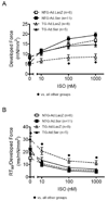
Familial hypertrophic cardiomyopathy (FHC) is an autosomal dominant genetic disorder linked to numerous mutations in the sarcomeric proteins. The clinical presentation of FHC is highly variable, but it is a major cause of sudden cardiac death in young adults with no specific treatments. We tested the hypothesis that early intervention in Ca(2+) regulation may prevent pathological hypertrophy and improve cardiac function in a FHC displaying increased myofilament sensitivity to Ca(2+) and diastolic dysfunction. A transgenic (TG) mouse model of FHC with a mutation in tropomyosin at position 180 was employed. Adenoviral-Serca2a (Ad.Ser) was injected into the left ventricle of 1-day-old non-transgenic (NTG) and TG mice. Ad.LacZ was injected as a control. Serca2a protein expression was significantly increased in NTG and TG hearts injected with Ad.Ser for up to 6 weeks. Compared to TG-Ad.LacZ hearts, the TG-Ad.Ser hearts showed improved whole heart morphology. Moreover, there was a significant decline in ANF and β-MHC expression. Developed force in isolated papillary muscle from 2- to 3-week-old TG-Ad.Ser hearts was higher and the response to isoproterenol (ISO) improved compared to TG-Ad.LacZ muscles. In situ hemodynamic measurements showed that by 3 months the TG-Ad.Ser hearts also had a significantly improved response to ISO compared to TG-Ad.LacZ hearts. The present study strongly suggests that Serca2a expression should be considered as a potential target for gene therapy in FHC. Moreover, our data imply that development of FHC can be successfully delayed if therapies are started shortly after birth.
Copyright © 2010 Elsevier Ltd. All rights reserved.
Figures


Similar articles
-
Rescue of tropomyosin-induced familial hypertrophic cardiomyopathy mice by transgenesis.Am J Physiol Heart Circ Physiol. 2007 Aug;293(2):H949-58. doi: 10.1152/ajpheart.01341.2006. Epub 2007 Apr 6. Am J Physiol Heart Circ Physiol. 2007. PMID: 17416600
-
Diastolic dysfunction in familial hypertrophic cardiomyopathy transgenic model mice.Cardiovasc Res. 2009 Apr 1;82(1):84-92. doi: 10.1093/cvr/cvp016. Epub 2009 Jan 15. Cardiovasc Res. 2009. PMID: 19150977 Free PMC article.
-
Functional effects of a tropomyosin mutation linked to FHC contribute to maladaptation during acidosis.J Mol Cell Cardiol. 2011 Mar;50(3):442-50. doi: 10.1016/j.yjmcc.2010.10.032. Epub 2010 Nov 1. J Mol Cell Cardiol. 2011. PMID: 21047515 Free PMC article.
-
[Lentiviral vector-mediated in vivo SERCA2a gene transfer improved cardiac function and remodeling of failing heart induced by myocardial infarction in the rat].Nihon Yakurigaku Zasshi. 2007 Apr;129(4):238-44. doi: 10.1254/fpj.129.238. Nihon Yakurigaku Zasshi. 2007. PMID: 17435333 Review. Japanese. No abstract available.
-
Sarcomeric proteins and familial hypertrophic cardiomyopathy: linking mutations in structural proteins to complex cardiovascular phenotypes.Heart Fail Rev. 2005 Sep;10(3):237-48. doi: 10.1007/s10741-005-5253-5. Heart Fail Rev. 2005. PMID: 16416046 Review.
Cited by
-
Molecular effects of the myosin activator omecamtiv mecarbil on contractile properties of skinned myocardium lacking cardiac myosin binding protein-C.J Mol Cell Cardiol. 2015 Aug;85:262-72. doi: 10.1016/j.yjmcc.2015.06.011. Epub 2015 Jun 20. J Mol Cell Cardiol. 2015. PMID: 26100051 Free PMC article.
-
Role of Genetics in Diagnosis and Management of Hypertrophic Cardiomyopathy: A Glimpse into the Future.Biomedicines. 2024 Mar 19;12(3):682. doi: 10.3390/biomedicines12030682. Biomedicines. 2024. PMID: 38540296 Free PMC article. Review.
-
Tropomyosin dephosphorylation results in compensated cardiac hypertrophy.J Biol Chem. 2012 Dec 28;287(53):44478-89. doi: 10.1074/jbc.M112.402040. Epub 2012 Nov 12. J Biol Chem. 2012. PMID: 23148217 Free PMC article.
-
Hypertrophic Cardiomyopathy: From Phenotype and Pathogenesis to Treatment.Front Cardiovasc Med. 2021 Oct 25;8:722340. doi: 10.3389/fcvm.2021.722340. eCollection 2021. Front Cardiovasc Med. 2021. PMID: 34760939 Free PMC article. Review.
-
PPP1R12C Promotes Atrial Hypocontractility in Atrial Fibrillation.Circ Res. 2023 Oct 13;133(9):758-771. doi: 10.1161/CIRCRESAHA.123.322516. Epub 2023 Sep 22. Circ Res. 2023. PMID: 37737016 Free PMC article.
References
Publication types
MeSH terms
Substances
Grants and funding
- R01 HL079032/HL/NHLBI NIH HHS/United States
- R01 HL81680/HL/NHLBI NIH HHS/United States
- R37 HL022231/HL/NHLBI NIH HHS/United States
- R01 HL088434/HL/NHLBI NIH HHS/United States
- R01 HL083156/HL/NHLBI NIH HHS/United States
- R01 HL078691/HL/NHLBI NIH HHS/United States
- P01 HL62426/HL/NHLBI NIH HHS/United States
- HL64035/HL/NHLBI NIH HHS/United States
- R01 HL071763/HL/NHLBI NIH HHS/United States
- R01HL22231/HL/NHLBI NIH HHS/United States
- R01 HL064035/HL/NHLBI NIH HHS/United States
- P01 HL062426/HL/NHLBI NIH HHS/United States
- HL80498/HL/NHLBI NIH HHS/United States
- HL71763/HL/NHLBI NIH HHS/United States
- R01 HL080498/HL/NHLBI NIH HHS/United States
- HL83156/HL/NHLBI NIH HHS/United States
- T32 HL07692/HL/NHLBI NIH HHS/United States
- T32 HL007692/HL/NHLBI NIH HHS/United States
- R01 HL081680/HL/NHLBI NIH HHS/United States
- R01 HL79032/HL/NHLBI NIH HHS/United States
LinkOut - more resources
Full Text Sources
Medical
Molecular Biology Databases
Research Materials
Miscellaneous

