

Prenatal L-DOPA exposure produces lasting changes in brain dopamine content, cocaine-induced dopamine release and cocaine conditioned place preference
- PMID: 20854831
- PMCID: PMC3014452
- DOI: 10.1016/j.neuropharm.2010.09.012
Prenatal L-DOPA exposure produces lasting changes in brain dopamine content, cocaine-induced dopamine release and cocaine conditioned place preference
Abstract
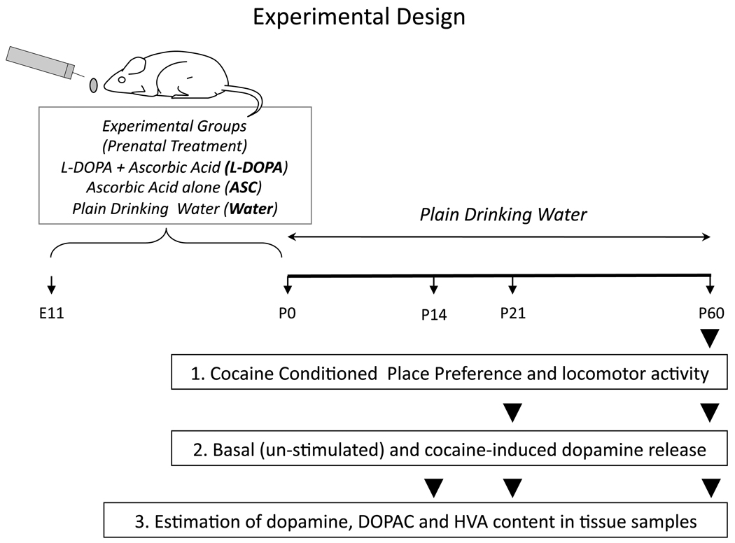
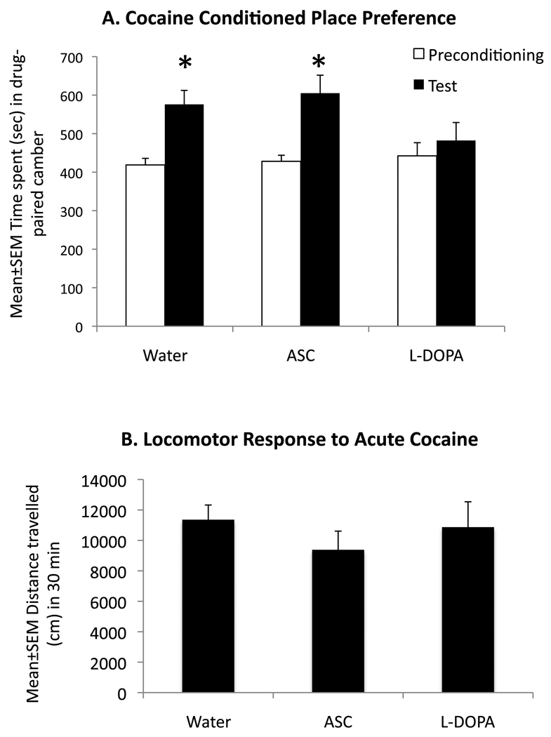
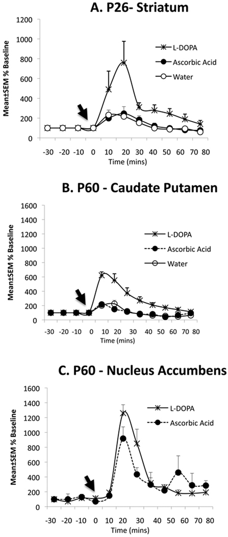
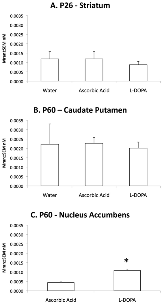
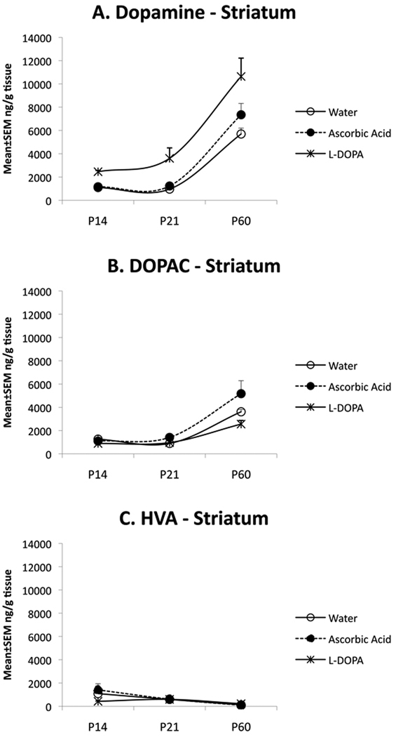
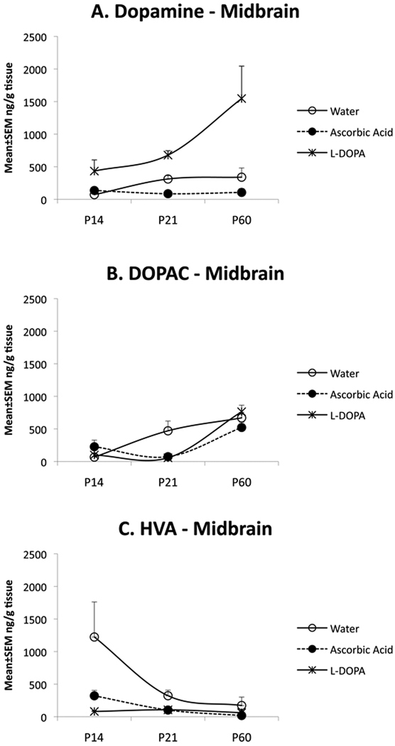
Dopamine, its receptors and transporter are present in the brain beginning from early in the embryonic period. Dopamine receptor activation can influence developmental events including neurogenesis, neuronal migration and differentiation raising the possibility that dopamine imbalance in the fetal brain can alter development of the brain and behavior. We examined whether elevated dopamine levels during gestation can produce persisting changes in brain dopamine content and dopamine-mediated behaviors. We administered L-3,4-dihydroxyphenylalanine (L-DOPA) in drinking water to timed-pregnant CD1 mice from the 11th day of gestation until the day of parturition. The prenatal L-DOPA exposure led to significantly lower cocaine conditioned place preference, a behavioral test of reward, at postnatal day 60 (P60). However, in vivo microdialysis measurements showed significant increases in cocaine-induced dopamine release in the caudate putamen of P26 and P60 mice exposed to L-DOPA prenatally, ruling out attenuated dopamine release in the caudate putamen as a contributor to decreased conditioned place preference. Although dopamine release was induced in the nucleus accumbens of prenatally L-DOPA exposed mice at P60 by cocaine, the dopamine release in the nucleus accumbens was not significantly different between the L-DOPA and control groups. However, basal dopamine release was significantly higher in the prenatally L-DOPA exposed mice at P60 suggesting that the L-DOPA exposed mice may require a higher dose of cocaine for induction of cocaine place preference than the controls. The prenatal L-DOPA exposure did not alter cocaine-induced locomotor response, suggesting dissociation between the effects of prenatal L-DOPA exposure on conditioned place preference and locomotor activity. Tissue concentration of dopamine and its metabolites in the striatum and ventral midbrain were significantly affected by the L-DOPA exposure as well as by developmental changes over the P14-P60 period. Thus, elevation of dopamine levels during gestation can produce persisting changes in brain dopamine content, cocaine-induced dopamine release and cocaine conditioned place preference.
Copyright © 2010 Elsevier Ltd. All rights reserved.
Figures
Similar articles
-
Effects of the plant-derived hallucinogen salvinorin A on basal dopamine levels in the caudate putamen and in a conditioned place aversion assay in mice: agonist actions at kappa opioid receptors.Psychopharmacology (Berl). 2005 May;179(3):551-8. doi: 10.1007/s00213-004-2087-0. Epub 2005 Jan 29. Psychopharmacology (Berl). 2005. PMID: 15682306
-
Augmentation of cocaine-sensitized dopamine release in the nucleus accumbens of adult mice following prenatal cocaine exposure.Dev Neurosci. 2009;31(1-2):76-89. doi: 10.1159/000207496. Epub 2009 Apr 17. Dev Neurosci. 2009. PMID: 19372689
-
The effects of prenatal cocaine, post-weaning housing and sex on conditioned place preference in adolescent rats.Psychopharmacology (Berl). 2014 Apr;231(8):1543-55. doi: 10.1007/s00213-013-3418-9. Epub 2014 Jan 17. Psychopharmacology (Berl). 2014. PMID: 24435324 Free PMC article.
-
Comparison of cocaine- and methamphetamine-evoked dopamine and glutamate overflow in somatodendritic and terminal field regions of the rat brain during acute, chronic, and early withdrawal conditions.Ann N Y Acad Sci. 2001 Jun;937:93-120. doi: 10.1111/j.1749-6632.2001.tb03560.x. Ann N Y Acad Sci. 2001. PMID: 11458542 Review.
-
Prenatal cocaine exposure alters signal transduction in the brain D1 dopamine receptor system.Ann N Y Acad Sci. 1998 Jun 21;846:238-47. Ann N Y Acad Sci. 1998. PMID: 9668411 Review.
Cited by
-
Electroacupuncture inhibition of hyperalgesia in rats with adjuvant arthritis: involvement of cannabinoid receptor 1 and dopamine receptor subtypes in striatum.Evid Based Complement Alternat Med. 2013;2013:393460. doi: 10.1155/2013/393460. Epub 2013 May 25. Evid Based Complement Alternat Med. 2013. PMID: 23762129 Free PMC article.
-
Placental Barrier and Autism Spectrum Disorders: The Roles of Prolactin and Dopamine in the Developing Fetal Brain-Part II.Innov Clin Neurosci. 2019 Nov 1;16(11-12):36-39. Innov Clin Neurosci. 2019. PMID: 32082942 Free PMC article.
-
Nicotine exposure of male mice produces behavioral impairment in multiple generations of descendants.PLoS Biol. 2018 Oct 16;16(10):e2006497. doi: 10.1371/journal.pbio.2006497. eCollection 2018 Oct. PLoS Biol. 2018. PMID: 30325916 Free PMC article.
-
Pre-gestational stress alters stress-response of pubertal offspring rat in sexually dimorphic and hemispherically asymmetric manner.BMC Neurosci. 2013 Jul 8;14:67. doi: 10.1186/1471-2202-14-67. BMC Neurosci. 2013. PMID: 23829597 Free PMC article.
-
Prenatal nicotine exposure mouse model showing hyperactivity, reduced cingulate cortex volume, reduced dopamine turnover, and responsiveness to oral methylphenidate treatment.J Neurosci. 2012 Jul 4;32(27):9410-8. doi: 10.1523/JNEUROSCI.1041-12.2012. J Neurosci. 2012. PMID: 22764249 Free PMC article.
References
-
- Bhide PG. Dopamine, cocaine and the development of cerebral cortical cytoarchitecture: A review of current concepts Seminars. Cell Dev. Biol. 2009;20:395–402. - PubMed
-
- Biederman J. Attention-deficit/hyperactivity disorder: a selective overview. Biol Psychiatry. 2005;57:1215–1220. - PubMed
-
- Cadoni C, Solinas M, Di Chiara G. Psychostimulant sensitization: differential changes in accumbal shell and core dopamine. Eur J Pharmacol. 2000;388:69–76. - PubMed
-
- Carelli RM. Nucleus accumbens cell firing and rapid dopamine signaling during goal-directed behaviors in rats. Neuropharmacology. 2004;47:180–189. - PubMed
Publication types
MeSH terms
Substances
Grants and funding
LinkOut - more resources
Full Text Sources
Other Literature Sources
Research Materials
