. 2010 Dec;42(12):2037-46.
doi: 10.1016/j.biocel.2010.09.008. Epub 2010 Sep 18.

N-terminal truncation of Stat5a/b circumvents PIAS3-mediated transcriptional inhibition of Stat5 in prostate cancer cells

Affiliations

N-terminal truncation of Stat5a/b circumvents PIAS3-mediated transcriptional inhibition of Stat5 in prostate cancer cells

Ayush Dagvadorj et al. Int J Biochem Cell Biol. 2010 Dec.

Abstract

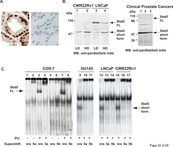
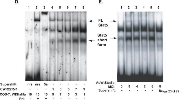
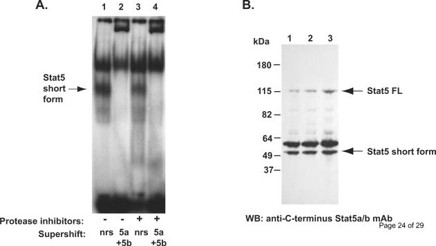
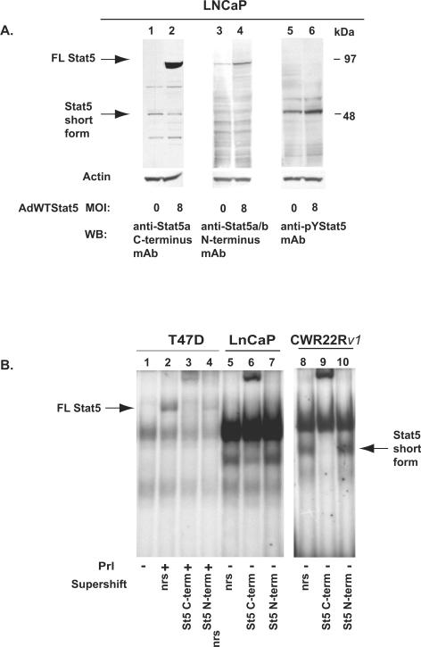
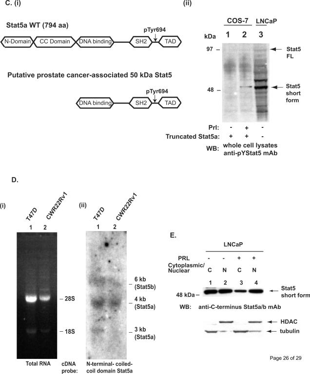
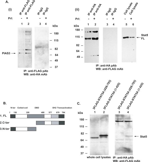
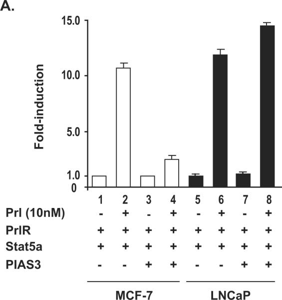
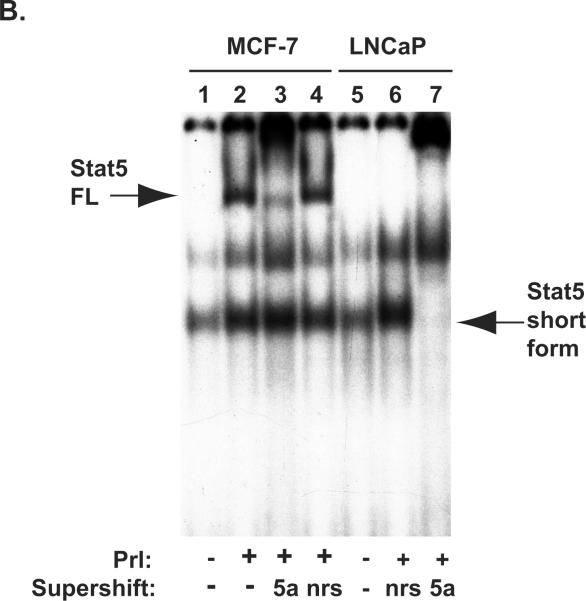
Transcription factor Stat5a/b is critical for prostate cancer cell survival and for prostate xenograft tumor growth. In addition, the Stat5a/b signaling pathway may contribute to progression of organ-confined prostate cancer to castration-resistant and/or metastatic disease. Expression of nuclear Stat5a/b is clustered to high grade human prostate cancers, and nuclear Stat5a/b in primary prostate cancer predicts early disease recurrence after initial treatment. Here, we show by Western blotting and electromobility shift assay that Stat5a/b protein in human prostate cancer is N-terminally truncated. This short form of Stat5a/b is generated post-translationally in vivo in prostate cancer cells and is the predominant form of Stat5a/b that binds to DNA. We further demonstrate by mutagenesis and co-immunoprecipitations that the N-domain of Stat5a/b is required for binding to PIAS3, and that PIAS3 inhibits transcriptional activity of Stat5a/b in breast cancer cells but not in prostate cancer cells. Thus, the proteolytic cleavage of the N-terminus of Stat5a/b may be a mechanism by which Stat5 evades the transcriptional repression by PIAS3 in prostate cancer cells, and results in increased Stat5-driven gene expression and prostate cancer progression.


Figures

Figure 1
Figure 1. Stat5a/b is cleaved to a truncated protein in human prostate cancer cells
A, Immunohistochemical detection of Stat5a/b in human prostate cancer using a monoclonal anti-pYStat5 antibody (left panel) with isotype-specific IgG as control (right panel). B, Western blotting of whole cell extracts of CWR22Rv1 and LNCaP cells grown at low density (LD) or high density (HD) and tissue extracts of three clinical human prostate cancers with anti-Stat5a/b mAb showing the full length (FL) and a truncated Stat5a/b (Stat5 short form) of approximately 50 kDa in size (arrows). C, EMSA of nuclear extracts of COS-7 cells transfected with Stat5a and stimulated with human prolactin (hPrl) using the Prl-response element of the beta–casein gene promoter as the probe (lanes 3, 4, 7 and 8). Stat5a and Stat5b complexes were supershifted with anti-Stat5a (lane 4) and anti-Stat5b (lane 8) pAbs indicating the specificity of the DNA-binding complexes. EMSA of nuclear extracts of human prostate cancer cell lines DU145 (lanes 9–11), LNCaP (lanes 12–14) and CWR22Rv1 (lanes 15–17) showing the presence of a smaller truncated form of Stat5a/b (Stat5 short form) which was supershifted by anti-Stat5a and anti-Stat5b pAbs. D, Nuclear extracts of COS-7 cells transfected with Stat5a, stimulated with hPrl for 30 min and supershifted with anti-Stat5a pAb (lanes 1–3) were mixed in various ratios with nuclear extracts from CWR22Rv1 cells and analyzed by EMSA (lanes 4–8). E, Full-length (FL) wild-type (Wt) Stat5a was introduced at decreasing doses (MOI 8, 4 and 2) to CWR22Rv1 cells using adenovirus as the expression vector (AdWtStat5a). EMSA shows the conversion of the full-length WtStat5a to the short form of Stat5a (the lower arrows on the left) (lanes 2–5). The short form Stat5a was supershifted by anti-Stat5a pAb (lane 6).
Figure 2
Figure 2. Short form STAT5 occurs naturally in prostate cancer cells
A, Electromobility shift assay of nuclear extract of DU145 cells prepared from flash frozen cell pellets in the absence (lane 1 and 2) or presence (lane 3 and 4) of protease inhibitors (phenylmethylsulfonylfluoride, aprotinin, leupeptin and pepstatin-A). The short form Stat5a/b (arrow) was detected in DU145 cells in the presence or in the absence of protease inhibitors, and the short form Stat5a/b was super-shifted by anti-Stat5a and anti-Stat5b antibodies. B, The short form Stat5a/b is not generated in vitro after disruption of the cell membranes. DU145 cell pellets were flash frozen in liquid nitrogen and directly lysed in boiling SDS loading buffer (lane 1), or the cell pellets were lysed in cell lysis buffer in the presence (lane 2) or in the absence of proteases inhibitors (lane 3) and boiled in SDS loading buffer and analyzed by Western blotting using anti-C-terminus Stat5a/b mAb.
Figure 3
Figure 3. The short form Stat5a/b expressed in human prostate cancer cells is N-terminally truncated and generated at posttranslational level
A, LNCaP cells were mock-infected or infected with AdWtStat5a at MOI of 8. Equal amounts of proteins were loaded in each lane, separated and immunoblotted with anti-Stat5a/b (lanes 1–2) mAbs raised against the C-terminal epitopes. The full-length (FL) and short Stat5a/b forms are indicated by arrows (left). Immunoblotting with anti-Stat5a/b mAb against epitopes in the N-terminus of Stat5a/b failed to recognize the short form of Stat5a/b in the parallel samples (lanes 3–4). Immunoblotting with the anti-pYStat5a/b mAb of parallel samples indicated that the majority of the N-terminally truncated short form Stat5a/b was phosphorylated. Actin immunoblotting demonstrates equal amounts of proteins loaded. B, EMSA of T47D human breast cancer cells stimulated with hPrl for 30 min with unstimulated cells as control show the full-length Stat5a/b expression in human breast cancer cells (arrow on left) (lanes 1–4) while not in human prostate cancer cells LNCaP (lanes 5–7) or CWR22Rv1 cells (lanes 8–10). The full-length Stat5a/b in T47D breast cancer cells was supershifted with the anti-Stat5a/b antibodies against the N-terminus or C-terminus of Stat5a/b (lanes 3 and 4), while the short form Stat5a/b in LNCaP and CWR22Rv1 cells (arrow on right) was not supershifted with only the antibody against the N-terminus of Stat5a/b (lanes 7 and 10). C, Schematic presentation of the truncated Stat5a generated by cloning (i). The truncated Stat5a lacking the N-terminal and coiled-coil (CC)-domain was transfected to COS-7 cells (ii). COS-7 cells were starved and stimulated with Prl (30 min) with unstimulated cells as control. Whole cell lysates of COS-7 cells were immunoblotted side-by-side with lysates of LNCaP cells with anti-pYStat5a/b mAb. D, Ethidium bromide staining of total RNA extracted from T47D human breast cancer cells or CWR22Rv1 prostate cancer cells and separated on 1% agarose gel (i). Hybridization of the Northern blot with a cDNA probe of the N-terminal and CC-domain of Stat5a demonstrates equal sizes of the mRNA transscripts in breast cancer cells expressing the full-length Stat5a protein and prostate cancer cells expressing the short form Stat5a/b protein (ii). E, Western blotting of cytoplasmic and nuclear (N) extracts of LNCaP cells with anti-C-terminus Stat5a/b mAb. Before preparation of the cytoplasmic and nuclear extracts, the cells had been starved for 16 h and stimulated with 10 nM human prolactin (Prl) for 30 min. Anti-HDAC and anti-tubulin immunoblotiing verify the quality of the nuclear and cytoplasmic extracts (lower panel).
Figure 4
Figure 4. PIAS physically interacts with the N-terminus of STAT5a
A, COS-7 cells were co-transfected with pFlag-Stat5a, pHA-PIAS3 and pPrlR, serum-starved for 16 h, then stimulated with 10 nM human Prl (Pr) for 30 min with unstimulated cells as control. Stat5a was immunoprecipitated with anti-FLAG pAb and blotted with anti-HA mAb (i). Stat5a formed a complex with PIAS3 (lanes 1 and 2). In the converse experiments, PIAS3 was immunoprecipitated with anti-HA pAb and the immunoprecipitations were blotted with anti-FLAG mAb (ii). B, Schematic presentation of the deletion constructs of Stat5a that were either C-terminally truncated or N-terminally truncated and tagged with the FLAG-epitopes. C, COS-7 cells were co-transfected with pFlag-Stat5a (426–793), pFlag-Stat5a (1–415) or pHA-PIAS3. PIAS3 was immunoprecipitated with anti-HA pAb and immunoblotted with anti-FLAG mAb recognizing the Flag-tagged N-terminally or C-terminally truncated Stat5a. The C-terminally truncated Stat5a (1–425) formed complexes with PIAS3 (lane 4) while the N-terminally truncated Stat5a (426–793) failed to co-immunoprecipitate with PIAS3 (lane 3).
Figure 5
Figure 5. Transcriptional activity of ligand-induced Stat5a in beta-Casein promoter-luciferase assay is repressed by PIAS3 in human breast cancer cells but not in human prostate cancer cells
A, MCF-7 cells and LNCaP cells (0.25 × 10⁶ cells/well) were transiently co-transfected with genomic beta-Casein-promoter-luciferase plasmid (0.5 μg), pRL-TK (0.025 μg), pPrl-receptor (PrlR) (0.25 μg), pStat5a (0.25 μg), and/or pPIAS3 (0.5 μg), as indicated. The total amount of plasmid DNA transfected was normalized to 1.275μg/well by addition of the empty vector (pMod-DNR). The cells were serum-starved for 20 h and treated for 16 h with (+) or without (−) 10 nM human hPrl as indicated. The mean values of three independent experiments performed in triplicates are presented, and S.E. values are indicated by bars. B, MCF-7 cells and LNCaP cells (1.5 × 10⁶ cells/dish) were transiently co-transfected with genomic pStat5a (2.0 μg) and pPrl-receptor (PrlR) (2.0 μg). The cells were serum-starved for 20 hr and treated with (+) or without (−) 10 nM human hPrl for 30 min. The nuclear extracts were prepared for EMSA assay. EMSA shows the presence of short form Stat5a in LNCaP cells but not in MCF-7 cells.

Similar articles

Cited by

References

    1. Abdulghani J, Gu L, Dagvadorj A, Lutz J, Leiby B, Bonuccelli G, et al. Stat3 promotes metastatic progression of prostate cancer. Am J Pathol. 2008;172:1717–28. - PMC - PubMed
    1. Ahonen TJ, Harkonen PL, Rui H, Nevalainen MT. PRL signal transduction in the epithelial compartment of rat prostate maintained as long-term organ cultures in vitro. Endocrinology. 2002;143:228–38. - PubMed
    1. Ahonen TJ, Xie J, LeBaron MJ, Zhu J, Nurmi M, Alanen K, et al. Inhibition of transcription factor Stat5 induces cell death of human prostate cancer cells. J Biol Chem. 2003;278:27287–92. - PubMed
    1. Assikis VJ, Do KA, Wen S, Wang X, Cho-Vega JH, Brisbay S, et al. Clinical and biomarker correlates of androgen-independent, locally aggressive prostate cancer with limited metastatic potential. Clin Cancer Res. 2004;10:6770–8. - PubMed
    1. Azam M, Lee C, Strehlow I, Schindler C. Functionally distinct isoforms of STAT5 are generated by protein processing. Immunity. 1997;6:691–701. - PubMed

Publication types

MeSH terms