

Emotion processing influences working memory circuits in pediatric bipolar disorder and attention-deficit/hyperactivity disorder
- PMID: 20855051
- PMCID: PMC2957818
- DOI: 10.1016/j.jaac.2010.07.009
Emotion processing influences working memory circuits in pediatric bipolar disorder and attention-deficit/hyperactivity disorder
Abstract
Objective: This functional magnetic resonance imaging (fMRI) study examined how working memory circuits are affected by face emotion processing in pediatric bipolar disorder (PBD) and attention-deficit/hyperactivity disorder (ADHD).
Methods: A total of 23 patients with PBD, 14 patients with ADHD, and 19 healthy control (HC) subjects (mean age, 13.36 ± 2.55 years) underwent an affective, two-back fMRI task with blocks of happy, angry, and neutral faces.
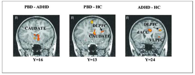
Results: For angry versus neutral faces PBD patients, relative to ADHD patients, exhibited increased activation in the subgenual anterior cingulate cortex (ACC) and orbitofrontal cortex, and reduced activation in the dorsolateral prefrontal cortex (DLPFC) and premotor cortex. Relative to the HC group, the PBD group showed no increased activation and reduced activation at the junction of DLPFC and ventrolateral prefrontal cortex (VLPFC). Relative to HC, the ADHD patients exhibited greater activation in the DLPFC and reduced activation in the ventral and medial PFC, pregenual ACC, striatum, and temporo-parietal regions. For happy versus neutral faces, relative to the ADHD group, the PBD group exhibited greater activation in the bilateral caudate, and relative to the HC group the ADHD group showed increased activation in the DLPFC, striatal, and parietal regions, and no reduced activation. The ADHD group, compared with the HC group, showed no reduced activation and increased activation in regions that were underactive for the angry face condition.
Conclusions: Relative to the ADHD group, the PBD group exhibited greater deployment of the emotion-processing circuitry and reduced deployment of working memory circuitry. Commonalities across PBD and ADHD patients, relative to the HC individuals, entailed cortico-subcortical activity that was reduced under negative emotional challenge and increased under positive emotional challenge.
Copyright © 2010 American Academy of Child and Adolescent Psychiatry. Published by Elsevier Inc. All rights reserved.
Figures


References
-
- Galanter CA, Leibenluft E. Frontiers between attention deficit hyperactivity disorder and bipolar disorder. Child and Adolescent Psychiatric Clinics of North America. 2008;17:325–346. - PubMed
-
- Insel TR, Cuthbert BN. Endophenotypes: bridging genomic complexity and disorder heterogeneity. Biological Psychiatry. 2009;66(11):988–989. - PubMed
-
- Geller B, Warner K, Williams M, Zimerman B. Prepubertal and young adolescent bipolarity versus ADHD: Assessment and validity using the WASH-U-KSADS, CBCL, and TRF. Journal of Affect Disorder. 1998;51(2):93–100. - PubMed
-
- Pavuluri MN, O’Connor MM, Harral EM, Sweeney JA. Affective neural circuitry during facial emotion processing in pediatric bipolar disorder. Biological Psychiatry. 2007;62(2):158–167. - PubMed
-
- Pavuluri MN, Passarotti AM. Neural bases of emotional processing in pediatric bipolar disorder. Expert Review of Neurotherapeutics. 2008;8(9):1381–1387. - PubMed
Publication types
MeSH terms
Grants and funding
LinkOut - more resources
Full Text Sources
Medical
Miscellaneous
