. 2010 Nov 25;407(2):333-40.
doi: 10.1016/j.virol.2010.08.019. Epub 2010 Sep 19.

Intrinsic disorder and oligomerization of the hepatitis delta virus antigen

Affiliations

Intrinsic disorder and oligomerization of the hepatitis delta virus antigen

Carolina Alves et al. Virology. .

Abstract

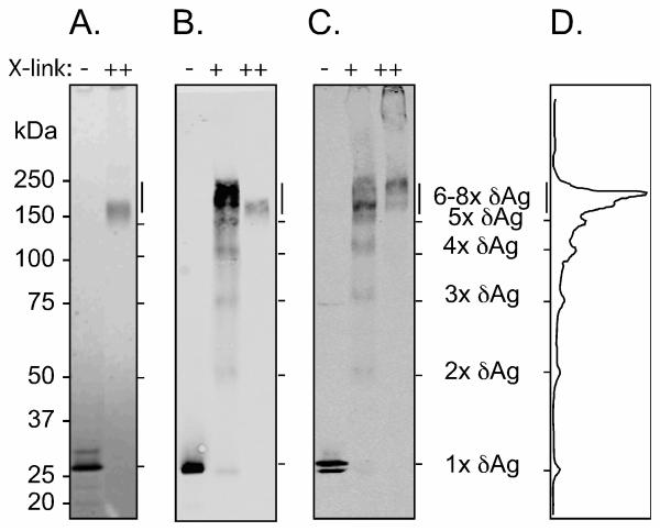
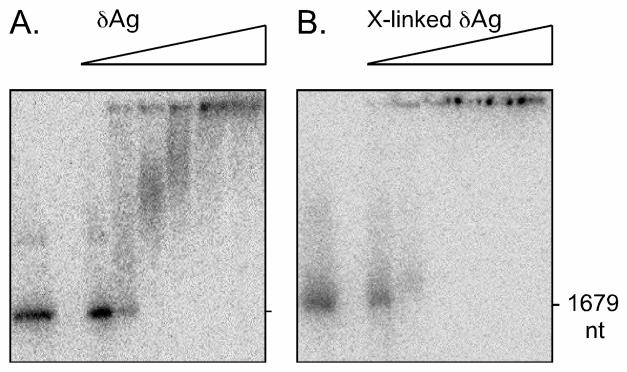
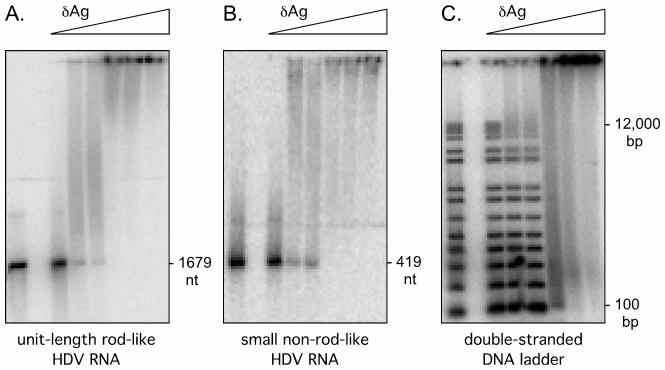
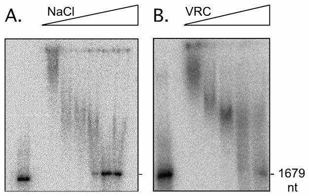
The 195 amino acid basic protein (δAg) of hepatitis delta virus (HDV) is essential for replication of the HDV RNA genome. Numerous properties have been mapped to full-length δAg and attempts made to link these to secondary, tertiary and quaternary structures. Here, for the full-size δAg, extensive intrinsic disorder was predicted using PONDR-FIT, a meta-predictor of intrinsic disorder, and evidenced by circular dichroism measurements. Most δAg amino acids are in disordered configurations with no more than 30% adopting an α-helical structure. In addition, dynamic light scattering studies indicated that purified δAg assembled into structures of as large as dodecamers. Cross-linking followed by denaturing polyacrylamide gel electrophoresis revealed hexamers to octamers for this purified δAg and at least this size for δAg found in virus-like particles. Oligomers of purified δAg were resistant to elevated NaCl and urea concentrations, and bound without specificity to RNA and single- and double-stranded DNAs.


Figures

Fig. 1
Fig. 1
Primary and secondary structure features of the 195 amino acid δAg. The upper panel indicates the coiled-coil domain (CDD), nuclear localization signal (NLS) and the RNA binding domain (RBD) (Han et al., 2009). The middle panel shows predictions of disorder using the meta-predictor PONDR-Fit (Xue et al., 2010), and three of its component programs (Obradovic et al., 2003; Obradovic et al., 2005), as indicated. The disorder score is a measure of the certainty that a region of the protein is disordered; a score of 1 indicates 100% certainty. The lower panel shows the primary sequence with basic, acidic and hydrophobic amino acids indicated in blue, red and green, respectively.
Fig. 2
Fig. 2
Predictions of δAg disorder for all 8 clades of HDV. Analyses were made as in Fig. 1. Also shown are controls of the highly disordered apoptin of chicken anemia virus, and the highly structured ammonia channel protein from E. coli.
Fig. 3
Fig. 3
Circular dichroism of δAg. The purified protein was analyzed as described in Materials and Methods. The open circles show the data points and the solid line the fit using the K2D2 program (Perez-Iratxeta and Andrade-Navarro, 2008) and the indicated results for α-helix, β-sheet and other (disordered) structures.
Fig. 4
Fig. 4
Electrophoretic mobility of purified δAg and of δAg assembled into virus-like particles. In panels A and B purified δAg, at 2 μM was treated without or with glutaraldehyde cross-linking, 0.01 or 0.1%, as indicated by + and ++ respectively. This was followed by SDS denaturation and electrophoresis on a gel of 4-12% polyacrylamide. In panel A, total protein was detected by SimplyBlue staining and in panel B, δAg was detected by immunoblot using specific antibody. In panel C, virus-like particles containing δAg, without and with prior cross-linking were examined as in panel B. The minor band migrating faster than the monomer might be a proteolytic fragment. At left are indicated MW markers. Panel D is the quantitation of panel B, lane 2. Indicated an interpretation of detected bands as multimers of δAg.
Fig. 5
Fig. 5
Electrophoretic mobility shift of HDV genomic unit-length RNA by δAg. Increasing amounts of δAg, without (panel A) or with (panel B) prior cross-linking with 0.1% glutaraldehyde followed by quenching with ammonium acetate, were incubated with trace amounts of 32P-labeled unit-length linear genomic HDV RNA. Samples were analyzed on non-denaturing gels of 1.5% agarose. After electrophoresis the gel was dried and radioactivity assayed using a bioimager. In each panel the first lane is the control in the absence of δAg. The triangle indicates use of increasing concentrations of δAg: 0.2, 0.4, 0.8, 1.6, 2 and 3.2 μM.
Fig. 6
Fig. 6
Electrophoretic mobility shift of RNA and DNA species by δAg. 32P-labeled RNA species were gel purified and then incubated in the absence or presence of increasing amounts of δAg. Samples were then subjected to electrophoresis under non-denaturing conditions, as in Fig. 5. In panel A the RNA was the 1679 nt unit-length antigenomic linear HDV RNA. In panel B it was a 419 nt species of antigenomic linear HDV RNA, representing the region 714-224, using the numbering of Kuo et al. (Kuo et al., 1988), that is predicted to not include the rod-like folding of the RNA. In panel C, we used end-labeled double-stranded DNA with a ladder of sizes from 100 to 12,000 bp. In each panel the left lane is the control in the absence of δAg. The triangles indicate use of increasing concentrations of δAg: 0.2, 0.4, 0.8, 1.6, 2 and 3.2 μM.
Fig. 7
Fig. 7
Binding of HDV RNA to δAg can be reversed by increasing concentrations of NaCl and VRC. 32P-labeled unit-length genomic HDV was incubated with δAg at 2 μM, and then increasing concentrations of NaCl (panel A) or VRC (panel B), and then analyzed as in Fig. 5. In each case the first lane is the control in the absence of δAg. The triangles indicate use of increasing concentrations of NaCl: 0.15, 0.3, 0.6, 1.2, 2.4, and 3.6 M (panel A) or VRC: 0, 0.3, 1, 3, and 10 mM (panel B).

Similar articles

Cited by

References

    1. Bergmann KF, Pohl C, Gerin JL. Characterization of proteins of hepatitis delta virus. Prog. Clin. Biol. Res. 1987;234:105–110. - PubMed
    1. Cao D, Haussecker D, Huang Y, Kay MA. Combined proteomic-RNAi screen for host factors involved in human hepatitis delta virus replication. RNA. 2009;15:1971–1979. - PMC - PubMed
    1. Casey JL. Hepatitis delta virus. In: Compans RM, Cooper MD, Honjo T, Koprowski H, Melchers F, Oldstone MBA, Olsnes S, Potter M, Vogt PK, Wagner H, editors. Current Topics in Microbiology and Immunology. Springer; Berlin: 2006.
    1. Chang FL, Chen PJ, Tu SJ, Chiu MN, Wang CJ, Chen DS. The large form of hepatitis δ antigen is crucial for the assembly of hepatitis d virus. Proc. Natl. Acad. Sci. USA. 1991;88:8490–8494. - PMC - PubMed
    1. Chang J, Gudima SO, Tarn C, Nie X, Taylor JM. Development of a novel system to study hepatitis delta virus genome replication. J. Virol. 2005;79:8182–8188. - PMC - PubMed

Publication types