Review
. 2010 Sep 20;190(6):953-63.
doi: 10.1083/jcb.201006020.

The cell biology of vision

Affiliations
Review

The cell biology of vision

Ching-Hwa Sung et al. J Cell Biol. .

Abstract

Humans possess the remarkable ability to perceive color, shape, and motion, and to differentiate between light intensities varied by over nine orders of magnitude. Phototransduction--the process in which absorbed photons are converted into electrical responses--is the first stage of visual processing, and occurs in the outer segment, the light-sensing organelle of the photoreceptor cell. Studies of genes linked to human inherited blindness have been crucial to understanding the biogenesis of the outer segment and membrane-trafficking of photoreceptors.


Figures

Figure 1.
Figure 1.
The visual sense organ. (A) Diagrams of the eye; an enlarged diagram of the fovea is shown in the box. Retina forms the inner lining of the most of the posterior part of the eye. The RPE is sandwiched between the retina and choroids, a vascularized and pigmented connective tissue. (B) Diagram of the organization of retinal cells. R, rod; C, cone; B, bipolar cell; H, horizontal cell; A, amacrine cell; G, ganglion cells; M, Müller cell. (C) An H&E-stained transverse section of human retina. Retina has laminated layers. The nuclei of the photoreceptors constitute the outer nuclear layer (ONL). The nuclei of the bipolar cells, amacrine cells, horizontal cells, and Müller glial cells are found in the inner nuclear layer (INL), and the nuclei of ganglion cells form the ganglion cell layer (GCL). The outer plexiform layer (OPL) contains the processes and synaptic terminals of photoreceptors, horizontal cells, and bipolar cells. The inner plexiform layer (IPL) contains the processes and terminals of bipolar cells, amacrine cells, and ganglion cells. The processes of Müller glial cells fill all space in the retina that is not occupied by neurons and blood vessels. Reproduced from Swaroop and Zack (2002), published by BioMed Central.
Figure 2.
Figure 2.
The morphological and molecular characteristics of vertebrate rod. (A) 3D cartoons depict the inter-relationship between rod and RPE (left) and IS–OS junction (right); RPE apical microvilli interdigitate the distal half of the OS. R, RPE; V, microvilli; O, OS; I, IS; N, nucleus, S, synaptic terminal. (B) A schematic drawing of a mammalian rod depicting its ciliary stalk and microtubule organizations; the axonemal (Ax) and cytoplasmic microtubules (not depicted) are anchored at the basal body in the distal IS. CP, calycal process; BB, basal body. The interactions between opposing membranes are depicted in color. The yellow shade indicates that the putative interaction of the ectodomains of usherin–VLGR1–whirlin complexes appear on both CC plasmalemma and the lateral plasmalemma of the IS ridge complex. The green shade indicates the putative chlosterol–prominin-1–protocadherin 21 interaction. (C) Electron micrographs reveal the hairpin loop structures of the disc rims and the fibrous links across the gap between the disc rims and plasma membranes (arrowheads). Bar, 100 nm. Reproduced from Townes-Anderson et al. (1988) with permission from J. Neurosci. (D) The OS plasma membrane and disc membrane have distinctive protein compositions; molecules are either expressed on the plasma membrane or the disc membranes, but not both. The only exception is rhodopsin; rhodopsin is present on disc membrane (with a much higher concentration) and plasma membrane (not depicted). The cGMP-gated channel: Na/Ca-K exchanger complex on the plasma membrane directly binds to the peripherin-2–ROM-1 oligomeric complex on the disc rim. The cGMP-gated channel is composed of three A1 subunits and one B1 subunit. ABCA4, a protein involved in retinoid cycle, is also enriched on the disc rim. RetGC1, retinal guanylyl cyclase; CNG channel, cGMP-gated channel. Adapted from Molday (2004). (E) Electron micrograph showing the longitudinal sectioning view of IS–OS junction of rat rod. Arrows point to the CC axonemal vesicles. An open arrow points to the fibrous structures linking the opposing membranes. Bar, 50 nm. Inset: a transverse section through the CC shows 9+0 arrangement; an arrow points to the cross-linker that gaps the microtubule doublet and adjacent ciliary membrane. R, apical IS ridge. Bar,100 nm. (F) Electron micrographs of a low-power (inset) and high-power images of the rat retina, at the junction between the rod OS and the RPE. MV, RPE microvillar processes enwrapped the distal OS. A white arrow points to a group of saccules from the tip of OS curls and upwards. White arrows in inset point to two distal OS fragments that are engulfed by RPE. Bar, 500 nm. Inset modified from Chuang et al. (2010) with permission from Mol. Biol. Cell.
Figure 3.
Figure 3.
OS morphology during normal rod development and in disease. (A) A drawing depicting the transformation of developing rods during their OS morphogenesis. (B) Representative electron micrograph of postnatal day 10 rat rods. This morphological appearance may represent a stage in OS morphogenesis. Many discs are longer than the matured discs; running in parallel to the ciliary stalk (unpublished data). Bar, 0.5 µm. (C) A rod from a postnatal day 15 mouse lacking RPGRIP1 (RPGRIP1−/−) containing vertically oriented discs is shown. Reproduced from Zhao et al. (2003), copyright The National Academy of Sciences, USA. Bar, 0.2 µm.
Figure 4.
Figure 4.
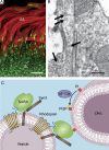
Vesicular targeting model explains how new discs are assembled at the base of the OS. (A) Rhodopsin is distributed evenly throughout the entire OS (red). In contrast, SARA (green) is specifically concentrated at the base of the OS and especially enriched on the vesicles residing in the basal 3-µm axonemal spaces. Bar, 5 µm. (B) Electron micrograph reveals the aberrant vesicle accumulation at the basal OS and at the CC shaft as a result of disruption of SARA function (arrows). Bar, 300 nm. (C) New discs at the base of the OS are assembled and “grow” via SARA-, PI3P-, and SNARE-mediated vesicular trafficking and membrane fusion. SARA directly binds to rhodopsin’s C terminus and syntaxin 3. The FYVE domain of SARA tethers axonemal vesicles to nascent discs through its high-affinity interaction with PI3P located on the immature discs. The close proximity of these membranes then permits the SNARE-mediated fusion event to happen. Panels A and B are reproduced from Chuang et al. (2007) with permission from Elsevier.
Figure 5.
Figure 5.
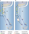
Vectorial OS trafficking of rhodopsin in ciliated photoreceptors. Photoreceptors possess two distinct populations of microtubules: axonemal and cytoplasmic. Both sets of microtubules have their minus ends anchored at the basal bodies (Troutt and Burnside, 1988). Cytoplasmic dynein 1 transports post-Golgi rhodopsin over long distances to reach the apical IS region. At least two current working models have been proposed to explain how rhodopsin is transported through the CC and how this process is coupled with rhodopsin’s disc incorporation. Panel A depicts a model in which four or more distinctive cellular processes are involved (Sedmak and Wolfrum, 2010). Step 1: Rhodopsin is fused with the apical IS plasmalemma, then crosses a putative structure barrier and reaches the ciliary membrane. Step 2: Rhodopsin is moved on the ciliary membrane using kinesin II–powered IFT. Step 3: Rhodopsin vesicle is internalized by the distal CC plasmalemma through endocytosis. Step 4: Rhodopsin vesicles undergo fusion to form nascent discs. Panel B depicts a model in which rhodopsin vesicles, which are generated in the IS, are delivered to the basal OS axoneme through channeling via the ciliary axonemal shaft. These vesicles then undergo fusion to form nascent discs. In this scenario, post-Golgi rhodopsin vesicles may traverse the endocytic compartments to recruit SARA and other elements for subsequent translocation and/or fusion.

References

    1. Abd-El-Barr M.M., Sykoudis K., Andrabi S., Eichers E.R., Pennesi M.E., Tan P.L., Wilson J.H., Katsanis N., Lupski J.R., Wu S.M. 2007. Impaired photoreceptor protein transport and synaptic transmission in a mouse model of Bardet-Biedl syndrome. Vision Res. 47:3394–3407 10.1016/j.visres.2007.09.016 - DOI - PMC - PubMed
    1. Andrews L.D., Cohen A.I. 1983. Freeze-fracture studies of photoreceptor membranes: new observations bearing upon the distribution of cholesterol. J. Cell Biol. 97:749–755 10.1083/jcb.97.3.749 - DOI - PMC - PubMed
    1. Avasthi P., Watt C.B., Williams D.S., Le Y.Z., Li S., Chen C.K., Marc R.E., Frederick J.M., Baehr W. 2009. Trafficking of membrane proteins to cone but not rod outer segments is dependent on heterotrimeric kinesin-II. J. Neurosci. 29:14287–14298 10.1523/JNEUROSCI.3976-09.2009 - DOI - PMC - PubMed
    1. Baccus S.A. 2007. Timing and computation in inner retinal circuitry. Annu. Rev. Physiol. 69:271–290 10.1146/annurev.physiol.69.120205.124451 - DOI - PubMed
    1. Baker S.A., Haeri M., Yoo P., Gospe S.M., III, Skiba N.P., Knox B.E., Arshavsky V.Y. 2008. The outer segment serves as a default destination for the trafficking of membrane proteins in photoreceptors. J. Cell Biol. 183:485–498 10.1083/jcb.200806009 - DOI - PMC - PubMed

Publication types