. 2010 Nov;30(22):5348-63.
doi: 10.1128/MCB.00353-09. Epub 2010 Sep 20.

Cooperative action of multiple cis-acting elements is required for N-myc expression in branchial arches: specific contribution of GATA3

Affiliations

Cooperative action of multiple cis-acting elements is required for N-myc expression in branchial arches: specific contribution of GATA3

Eric Potvin et al. Mol Cell Biol. 2010 Nov.

Erratum in

  • Mol Cell Biol. 2012 Dec;32(24):5152-3

Abstract

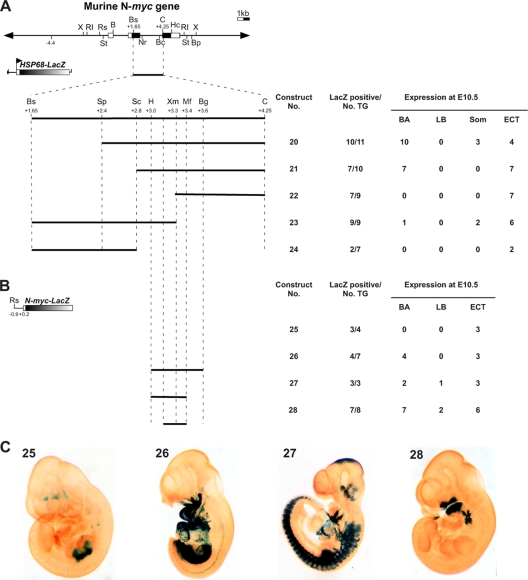
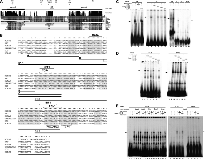
The precise expression of the N-myc proto-oncogene is essential for normal mammalian development, whereas altered N-myc gene regulation is known to be a determinant factor in tumor formation. Using transgenic mouse embryos, we show that N-myc sequences from kb -8.7 to kb +7.2 are sufficient to reproduce the N-myc embryonic expression profile in developing branchial arches and limb buds. These sequences encompass several regulatory elements dispersed throughout the N-myc locus, including an upstream limb bud enhancer, a downstream somite enhancer, a branchial arch enhancer in the second intron, and a negative regulatory element in the first intron. N-myc expression in the limb buds is under the dominant control of the limb bud enhancer. The expression in the branchial arches necessitates the interplay of three regulatory domains. The branchial arch enhancer cooperates with the somite enhancer region to prevent an inhibitory activity contained in the first intron. The characterization of the branchial arch enhancer has revealed a specific role of the transcription factor GATA3 in the regulation of N-myc expression. Together, these data demonstrate that correct N-myc developmental expression is achieved via cooperation of multiple positive and negative regulatory elements.


Figures

FIG. 1.
FIG. 1.
Expression profile of N-myc at E10.5. Whole-mount in situ hybridization was performed on E10.5 wild-type embryos (A to C, E, and F) and on E12.5 lung tissue (D). N-myc expression was detected in the central nervous system (A), in tissues derived from the neural crest, such as the dorsal root ganglia (B) and the branchial arches (C and F), and in somites (B) and limb buds (E). In branchial arches, N-myc expression was detected in the mesenchymal and ectodermal components, as shown in embryo sections from whole-mount in situ hybridization experiments and observed by interferential contrast microscopy (F). N-myc is highly expressed in lung epithelium (D), as previously described (42). DRG, dorsal root ganglia; ec, ectoderm; H, heart; Hy, hyoid arch; M, mesencephalon; Md, mandibular arch; me, mesenchyme; Mx, maxillary arch; Nt, neural tube; Som, somites; T, telencephalon.
FIG. 2.
FIG. 2.
Identification of a limb bud enhancer in upstream sequences of the mouse N-myc gene. (A) Structure of the mouse N-myc gene from which the various constructs were derived and diagram of the N-myc-lacZ transgenes used to generate F0 transgenic embryos. Open boxes indicate transcribed regions, and black boxes correspond to the translated sequences. The restriction enzymes used to isolate the fragments microinjected into fertilized eggs are indicated at each extremity. When two restriction enzymes sites are indicated, the upper one is unique in the construct and was used for the preparation of the fragment. B, BamHI; Bc, BclI; Bp, BspHI; Bs, BstBI; C, ClaI; Hc, HincII; N, NotI; Nr, NruI; RI, EcoRI; Rs, RsrII; S, SalI; St, StuI; X, XbaI. (B) Summary of the transgenic expression analysis. The number of lacZ-expressing embryos from the total number of transgenic embryos (TG) obtained is presented as well as the number of positively stained embryos for each structure listed. Som, somites; ECT, ectopic. (C) Representative X-Gal staining pattern for E10.5 transgenic embryos, showing the effects of the different 5′ deletions on lacZ expression. The number of the construct tested is identified in the upper left corner of each photo.
FIG. 3.
FIG. 3.
Downstream sequences of the N-myc gene are required for branchial arch expression and contain a somite enhancer. (A) Schematic representation of the N-myc-lacZ 3′ deletion transgenes used to generate F0 transgenic embryos. B, BamHI; Bc, BclI; Bp, BspHI; Bs, BstBI; C, ClaI; Hc, HincII; Nr, NruI; RI, EcoRI; Rs, RsrII; S, SalI; St, StuI; X, XbaI. (B) Summary of the transgenic expression analysis. TG, transgenic; Som, somites; ECT, ectopic. (C) Representative X-Gal staining obtained for E10.5 transgenic embryos showing the effects of the different 3′ deletions on lacZ expression. The construct number tested is identified in the upper left corner of each photo.
FIG. 4.
FIG. 4.
The second exon-intron of N-myc acts as a branchial arch enhancer but is not sufficient to direct branchial arch expression in the N-myc context. (A) Schematic representation of N-myc-lacZ intragenic deletion constructs used to generate F0 transgenic embryos. B, BamHI; Bc, BclI; Bp, BspHI; Bs, BstBI; C, ClaI; Hc, HincII; N, NotI; Nr, NruI; RI, EcoRI; Rs, RsrII; S, SalI; St, StuI; X, XbaI. (B) Summary of the transgenic expression analysis. TG, transgenic; Som, somites; ECT, ectopic. (C) Representative X-Gal staining obtained for E10.5 transgenic embryos, showing the effects of the different intragenic deletions on the lacZ expression pattern. The construct number is identified in the upper left corner of each photo.
FIG. 5.
FIG. 5.
Localization of the BA enhancer in the N-myc second intron. (A) Schematic representation of N-myc exon 2-intron 2 deletions tested with the hsp68-lacZ reporter in F0 transgenic embryos and summary of the transgenic expression analysis. B, BamHI; Bc, BclI; Bp, BspHI; Bs, BstBI; C, ClaI; Hc, HincII; Nr, NruI; RI, EcoRI; Rs, RsrII; St, StuI; X, XbaI; H, HindIII; Mf, MfeI; Sc, ScaI; Sp, SphI; Xm, XmnI. (B) Schematic representation of N-myc intron 2 sequences tested on a minimal N-myc promoter-lacZ reporter in F0 transgenic embryos and summary of the transgenic expression analysis. TG, transgenic; Som, somites; ECT, ectopic. (C) Representative X-Gal staining obtained for E10.5 transgenic embryos, showing the effects of the different intron 2 sequences for which results are shown in panel B, on the lacZ expression pattern. The construct is identified in the upper left corner of each photo.
FIG. 6.
FIG. 6.
Targeted expression of the lacZ reporter constructs in the developing BA. Transgenic embryos produced by the different N-myc-lacZ constructs were embedded in paraffin and sectioned to show in more detail the staining patterns. (A and B) Embryos derived from constructs 1 and 8, respectively, which included the promoter and the intragenic sequences of N-myc. X-Gal staining in the BA was detected in the mesenchyme (arrow) and in the ectoderm (arrowhead). (C and D) Embryos derived from constructs containing the N-myc minimal promoter and the 600-bp (C; construct 26) or 230-bp (D; construct 28) BA enhancer. X-Gal staining was mainly ectodermal in the specimen in panel C and mesenchymal in the specimen in panel D.
FIG. 7.
FIG. 7.
The 230-pb BA enhancer contains a consensus GATA binding motif. (A) Nucleotide sequence conservation in the N-myc second intron. A genomic sequence comparison of the mouse N-myc second intron with the genome of several species was performed using the UCSC genome browser. The regions with vertical lines indicate sequence conservation. The uppermost line shows a summary of conservation among species. The positions of the second and third exons of N-myc are indicated by black boxes, and the 230-pb BA enhancer is shown by the gray box labeled S1. (B) Alignment of the nucleotide sequences of the 230-bp BA enhancer among mammalian species. Asterisks indicate nucleotides conserved in all six species. Gray boxes indicate sequences with more than five consecutively conserved nucleotides. The transcription factor binding sites identified in a genome-wide computational prediction of transcriptional regulatory modules in mouse and human N-myc and corresponding to the sequences conserved in all six species are indicated http://genomequebec.mcgill.ca/PReMod (6, 16). Overlapping S1 subfragments (S1.1, S1.2, and S1.3) as well as subfragments of S1.1 (Oligos A, B, and C) used in the EMSA are indicated by lines under the sequence. (C) In vitro detection of protein binding to S1 sequences. EMSA with E10.5 protein extract were performed with S1 fragment (lanes 1 to 12) and subfragments (S1.1, S1.2 and S1.3; lanes 13 to 18) as probes. Specific binding complexes to S1 were assessed by the addition of a 100-fold excess of unlabeled S1 (lane 3), pBluescript sequences (lanes 4 and 10), S1.1 (lane 7), S1.2 (lane 8), or S1.3 (lane 9). (D) EMSA with S1.1 subfragments S1.1B and S1.1C. Specific binding complexes to subfragments S1.1B and S1.1C were assessed by competition with an excess of S1.1A (lanes 3 and 10), S1.1B (lanes 4 and 11), S1.1B* (fragment carrying a point mutation in the GATA consensus sequence binding site; lanes 5 and 12), S1.1AB (lanes 6 and 14), and S1.1C fragments (lanes 7 and 13). (E) EMSA with recombinant GATA proteins (GATA1, -3, -4, -5, and -6) and the S1.1B probe (lanes 3, 6, 9, 12, 15, and 20) or S1.1B* probe (lane 25). The specificity of binding was assessed by competition with an excess of S1.1A (lanes 5, 8, 11, 14, 17, 21, and 26) or S1.1B (lanes 4, 7, 10, 13, 16, 22, and 27) subfragments. Comp, competitor.
FIG. 8.
FIG. 8.
GATA binds to the BA enhancer in vivo and can activate transcription in trans-activation assays. (A) In a transient-transfection assay, mouse GATA1, -3, and -5 or rat GATA4 and -6 proteins upregulated transcription of a luciferase reporter construct containing the 600-bp intronic BA enhancer (BA/enh-pGL3) by a factor of 1.2 to 3.2. Values shown represent the means of relative luciferase activity obtained from six independent experiments ± the SEM. Statistical analyses using the t test were performed to determine whether the differences were statistically significant. *, P < 0.05; **, P < 0.005; ****, P < 0.00005. (B) NBA2 neuroblastoma cell cultures were cross-linked and chromatin was immunoprecipitated with mouse IgG, anti-GATA3 antibody, or anti-histone H3. Recruitment of GATA3 and H3 on four contiguous regions of the N-myc locus was evaluated by quantitative PCR and is indicated as the percentage of chromatin immunoprecipitation. The data are means ± SEM of three independent experiments.
FIG. 9.
FIG. 9.
The GATA binding site in the 230-bp BA enhancer is essential for the enhancer activity. (A) Schematic representation of N-myc-lacZ transgenes used to generate F0 transgenic embryos. The 230-bp BA enhancer was deleted in construct 29, whereas point mutations were introduced in the GATA binding site in construct 30 (GATA to TCGC). B, BamHI; Bc, BclI; Bp, BspHI; Bs, BstBI; C, ClaI; Hc, HincII; Nr, NruI; RI, EcoRI; Rs, RsrII; St, StuI; X, XbaI; N, NotI; S, SalI. (B) Summary of the transgenic expression analysis. No lacZ expression was observed in the BA for either construct. TG, transgenic; ECT, ectopic. (C) Representative X-Gal staining obtained for E10.5 transgenic embryos. DNA extracted from the yolk sacs was amplified by PCR to confirm the presence of the deletion or the point mutations in the GATA site. Constructs 1, 29, and 30 were amplified as controls. The point mutations in the GATA site introduced a new BbvI restriction site that was used to distinguish the endogenous gene (end) from the transgene (Tg) after restriction digestion. The expected bands for the endogenous gene and the transgene are indicated. d, BbvI digested; nd, not digested. (D) Reduced N-myc-lacZ transgene expression in Gata3/ embryos. To investigate the role of the GATA3 transcription factor in N-myc gene regulation, Gata3+/ and Gata3+/ Tg+/N-myc 41S mice were intercrossed. E9.25 embryos were recovered and stained for β-galactosidase activity in five independent experiments. As N-myc expression is very dynamic, the matings were performed over a 1-h period, and embryos at the same somite stage (±1 somite) were used for comparisons. Representative X-Gal staining showing the reduced N-myc-lacZ transgene expression in BA in the absence of GATA3 is presented. The specimens shown were embedded in paraffin and sectioned to show the X-Gal staining pattern in BA. Strong X-Gal staining was detected in the Gata3+/+ Tg+/N-myc 41S specimen, whereas it was barely detectable in Gata3/ Tg+/N-myc 41S BA specimens (arrowheads).
FIG. 10.
FIG. 10.
Model for the regulation of N-myc expression in branchial arches, somites (Som), and limb buds. N-myc branchial arch expression involves at least three regulatory domains, identified as R2, R3, and R4. The R3 domain corresponds to the second intron of N-myc and acts as a branchial arch enhancer when linked to a heterologous promoter. However, in the N-myc gene context, R3 requires the R4 domain, localized in 3′ sequences of the N-myc gene. The R4 domain is dispensable when the R2 domain, which corresponds to part of the first exon and the first intron, is deleted. Thus, the R3 branchial arch enhancer and the R4 branchial arch domain cooperate to overcome the inhibitory action of the R2 domain. The R4 domain also contains a somite enhancer, which also seems to be under the negative control of elements located in the R2 and the promoter regions of N-myc (not shown). Limb bud expression is under the control of a specific enhancer located upstream of the N-myc gene and identified as R1. B, BamHI; Bs, BspHI; C, ClaI; Hc, HincII; X, XbaI.

References

    1. Alt, F. W., R. DePinho, K. Zimmerman, E. Legouy, K. Hatton, P. Ferrier, A. Tesfaye, G. Yancopoulos, and P. Nisen. 1986. The human myc gene family. Cold Spring Harbor Symp. Quant. Biol. 51:931-941. - PubMed
    1. Aubry, S., and J. Charron. 2000. N-Myc shares cellular functions wih c-Myc. DNA Cell Biol. 19:353-364. - PubMed
    1. Babiss, L. E., and J. M. Friedman. 1990. Regulation of N-myc gene expression: use of an adenovirus vector to demonstrate posttranscriptional control. Mol. Cell. Biol. 10:6700-6708. - PMC - PubMed
    1. Baena, E., M. Ortiz, A. C. Martinez, and I. M. de Alboran. 2007. c-Myc is essential for hematopoietic stem cell differentiation and regulates Lin(-)Sca-1(+)c-Kit(-) cell generation through p21. Exp. Hematol. 35:1333-1343. - PubMed
    1. Ball, N. S., and P. H. King. 1997. Neuron-specific Hel-N1 and HuD as novel molecular markers of neuroblastoma: a correlation of HuD messenger RNA levels with favorable prognostic features. Clin. Cancer Res. 3:1859-1865. - PubMed

Publication types

MeSH terms