. 2010 Oct 5;107(40):17146-51.
doi: 10.1073/pnas.1004785107. Epub 2010 Sep 20.

Defining the mechanism of polymerization in the serpinopathies

Affiliations

Defining the mechanism of polymerization in the serpinopathies

Ugo I Ekeowa et al. Proc Natl Acad Sci U S A. .

Abstract

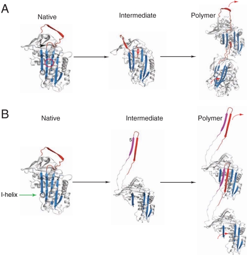
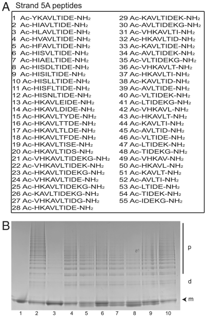
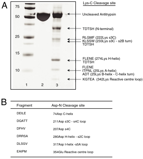
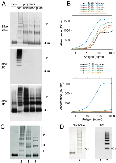
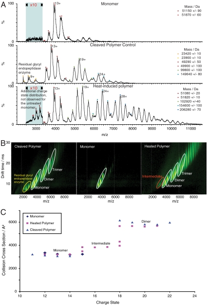
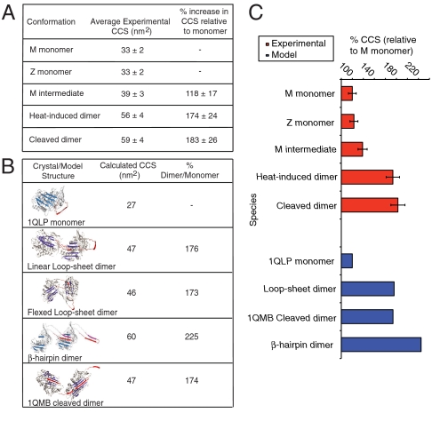
The serpinopathies result from the ordered polymerization of mutants of members of the serine proteinase inhibitor (serpin) superfamily. These polymers are retained within the cell of synthesis where they cause a toxic gain of function. The serpinopathies are exemplified by inclusions that form with the common severe Z mutant of α(1)-antitrypsin that are associated with liver cirrhosis. There is considerable controversy as to the pathway of serpin polymerization and the structure of pathogenic polymers that cause disease. We have used synthetic peptides, limited proteolysis, monoclonal antibodies, and ion mobility-mass spectrometry to characterize the polymerogenic intermediate and pathological polymers formed by Z α(1)-antitrypsin. Our data are best explained by a model in which polymers form through a single intermediate and with a reactive center loop-β-sheet A linkage. Our data are not compatible with the recent model in which polymers are linked by a β-hairpin of the reactive center loop and strand 5A. Understanding the structure of the serpin polymer is essential for rational drug design strategies that aim to block polymerization and so treat α(1)-antitrypsin deficiency and the serpinopathies.


Conflict of interest statement

The authors declare no conflict of interest.

Figures

Fig. 1.
Fig. 1.
The two proposed pathways of serpin polymerization. (A) The classical pathway of polymerization. The Z mutation (Glu342Lys-arrow head) or shutter domain mutations (red circle) destabilize β-sheet A (blue) to form an activated intermediate species (M*). Intermolecular linkage occurs via donation of the reactive loop (red) from one molecule to the open lower portion of the central β-sheet A channel of a second molecule. (B) The alternative β-hairpin pathway. The formation of an intermediate requires unraveling of the helix I (green arrow) and the extrusion of strand 5A (purple) from β-sheet A to generate a donor interface with the reactive center loop. Linkage occurs by the insertion of this hairpin domain into β-sheet A of a similarly activated molecule. The strands of β-sheet A are labeled.
Fig. 2.
Fig. 2.
Assessing the ability of strand 5A peptide analogues to inhibit the heat-induced polymerization of Z α1-antitrypsin. (A) The sequences of the 55 strand 5A analogue peptides assessed for their ability to block polymerization. The strand 5A sequence is KAVHKAVLTIDE. (B) 7.5% (w/v) acrylamide nondenaturing PAGE to evaluate the ability of strand 5A peptides to block the polymerization of Z α1-antitrypsin. The peptides were incubated at a molar ratio of 100∶1 with monomeric Z α1-antitrypsin (0.2 mg/mL) in PBS at 37 °C for 2 d. The temperature of the mixture was then raised to 41 °C for 5 d to form polymers. Lane 1: monomeric Z α1-antitrypsin; lane 2: control Z α1-antitrypsin polymers; lane 3: Z α1-antitrypsin incubated with the reactive loop blocking peptide TTAI; lanes 4–10: Z α1-antitrypsin incubated with strand 5A peptides (peptides 1–7 from A). The monomer (m), dimer (d) and polymer (p) are indicated. Each lane contains 4 μg of protein.
Fig. 3.
Fig. 3.
Endopeptidase cleavage of polymeric Z α1-antitrypsin. (A) 12% (w/v) SDS-PAGE to show the cleavage products of Lys-C digestion of polymeric Z α1-antitrypsin and their secondary structural location in the monomer. Lane 1: molecular mass markers (kDa); lane 2: undigested Z α1-antitrypsin; lane 3: Z α1-antitrypsin polymers incubated overnight with Lys-C. The gel was blotted onto PVDF and subjected to N-terminal sequencing. Longer incubation times did not change the cleavage profile. (B) Secondary structural location of cleavage fragments following Asp-N digestion of Z α1-antitrypsin polymers.
Fig. 4.
Fig. 4.
Monoclonal antibody 2C1 recognizes polymers of α1-antitrypsin prepared by heating but not by other denaturing conditions. (A) 7.5% (w/v) nondenaturing PAGE to show polymer analyzed by silver stain (upper) or Western blot, first with mAb 2C1 (center) and then with mAb 2D1 (bottom) on the same membrane. mAb 2C1 recognized heat-induced polymers but gave no signal for polymers prepared by treating α1-antitrypsin at low pH, or with 1–3 M guanidine or 1–4 M urea. The monomer (m) and polymer (p) are indicated. (B) The same polymers assayed in sandwich ELISA, using either mAb 9C5 (upper graph) or 2C1 (lower graph) as the detecting antibodies. mAb 9C5 detected all species with high affinity but mAb 2C1 detected only heat-induced polymers. (C) 7.5% (w/v) nondenaturing PAGE. Lane 1: M α1-antitrypsin monomer; lane 2: heat-induced α1-antitrypsin polymer; lane 3: antithrombin hexapeptide-induced polymer of α1-antitrypsin; lane 4: P9-P10 reactive center loop-cleaved polymer of α1-antitrypsin. The monomer (m), dimer (d), polymer (p) and intermediate state (I) are indicated. The intermediate in lane 2 migrates between the monomeric and dimeric states of the non heat-induced polymers. (D) 7.5% (w/v) nondenaturing PAGE of Z α1-antitrypsin monomer (lane 1) and polymer (lane 2) visualized by SimplyBlue™ (left) or following Western blot analysis with mAb 2C1 (right). Arrowheads indicate the intermediate (I) in the polymer lanes. The 2C1 shows no signal for the monomer but recognizes the intermediate and higher order polymers.
Fig. 5.
Fig. 5.
α1-antitrypsin polymers studied by IM-MS. (A) Mass spectra of M α1-antitrypsin monomer, the heat-induced polymer, and P9-P10 reactive loop-cleaved polymer. The mass spectrum of the monomer has a charge state distribution centered on the 13+ ion (upper spectrum). The mass spectrum of the reactive loop-cleaved polymer has four charge state distributions (middle spectrum). The peaks at low m/z are attributed to residual enzyme and the series of peaks at 4,000 and 5,250 and 6,250 m/z represent the cleaved monomer, dimer and trimer respectively. Similar spectra were observed for the heat-induced sample (lower spectrum). However, this spectrum exhibits a second monomer population with higher charge states (15+ to 18+), which are consistent with the polymerogenic intermediate. (B) Ion mobility drift times observed for the M α1-antitrypsin monomer, the heat-induced polymer, and reactive loop-cleaved polymer (corresponding mass spectra shown in A). The monomer and polymer species are labeled and highlighted with white ellipses, the intermediate with a red ellipse, and the cleavage enzyme with a yellow ellipse. (C) Drift times from (B) converted into a collision cross sectional plot. The monomer collision cross sections for all the samples are almost identical and similarly the collision cross sections for the cleaved polymer and heat-induced polymer dimers also overlap. The intermediate in the heat-induced polymer sample shows an expanded collision cross section relative to the monomer and maintains the same electrophoretic relationship with the dimer as that observed on nondenaturing PAGE (Fig. 4C).
Fig. 6.
Fig. 6.
Comparison of experimental and calculated collision cross sections (CCS) for models of α1-antitrypsin polymers. (A) Experimental collision cross sections for α1-antitrypsin monomers, the heat-induced and reactive loop-cleaved M α1-antitrypsin dimers. The dimer collision cross sections are indistinguishable. (B) Table of calculated collision cross section measurements for the X-ray crystallographic structures of monomeric α1-antitrypsin (1QLP), the cleaved dimer (1QMB), and the three-dimensional dimer models for the loop-sheet and β-hairpin pathways. The flexed loop-sheet, linear loop-sheet dimers and 1QMB cleaved dimers collision cross sections are very similar, whereas the β-hairpin dimer has a significantly larger collision cross section. (C) Plot comparing the percentage increases in collision cross section upon dimer formation for experimental (red) and calculated (blue) data. The relationship between the experimental heat-induced and reactive loop-cleaved dimer collision cross sections are consistent with a loop-sheet linkage and the crystallographic structure of cleaved dimers (1QMB). The calculated collision cross section of the β-hairpin dimer model is significantly larger than that observed experimentally for dimers formed by heating α1-antitrypsin.

References

    1. Gooptu B, Lomas DA. Conformational pathology of the serpins: themes, variations and therapeutic strategies. Annu Rev Biochem. 2009;78:147–176. - PubMed
    1. Sveger T. The natural history of liver disease in α1-antitrypsin deficient children. Acta Paediatr Scand. 1988;77:847–851. - PubMed
    1. Lomas DA, Evans DL, Finch JT, Carrell RW. The mechanism of Z α1-antitrypsin accumulation in the liver. Nature. 1992;357:605–607. - PubMed
    1. James EL, Bottomley SP. The mechanism of α1-antitrypsin polymerization probed by fluorescence spectroscopy. Arch Biochem Biophys. 1998;356:296–300. - PubMed
    1. Dafforn TR, Mahadeva R, Elliott PR, Sivasothy P, Lomas DA. A kinetic mechanism for the polymerization of α1-antitrypsin. J Biol Chem. 1999;274:9548–9555. - PubMed

Publication types

LinkOut - more resources