. 2010 Oct 5;107(40):17327-32.
doi: 10.1073/pnas.1008151107. Epub 2010 Sep 20.

Complement receptor 1 is the host erythrocyte receptor for Plasmodium falciparum PfRh4 invasion ligand

Affiliations

Complement receptor 1 is the host erythrocyte receptor for Plasmodium falciparum PfRh4 invasion ligand

Wai-Hong Tham et al. Proc Natl Acad Sci U S A. .

Abstract

Plasmodium falciparum is responsible for the most severe form of malaria disease in humans, causing more than 1 million deaths each year. As an obligate intracellular parasite, P. falciparum's ability to invade erythrocytes is essential for its survival within the human host. P. falciparum invades erythrocytes using multiple host receptor-parasite ligand interactions known as invasion pathways. Here we show that CR1 is the host erythrocyte receptor for PfRh4, a major P. falciparum ligand essential for sialic acid-independent invasion. PfRh4 and CR1 interact directly, with a K(d) of 2.9 μM. PfRh4 binding is strongly correlated with the CR1 level on the erythrocyte surface. Parasite invasion via sialic acid-independent pathways is reduced in low-CR1 erythrocytes due to limited availability of this receptor on the surface. Furthermore, soluble CR1 can competitively block binding of PfRh4 to the erythrocyte surface and specifically inhibit sialic acid-independent parasite invasion. These results demonstrate that CR1 is an erythrocyte receptor used by the parasite ligand PfRh4 for P. falciparum invasion.


Conflict of interest statement

The authors declare no conflict of interest.

Figures
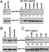
Fig. 1.
Fig. 1.
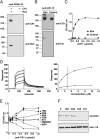
Anti-CR1 antibodies and sCR1 inhibit PfRh4 erythrocyte binding. (A) Anti-CR1 antibody (E11) inhibits PfRh4 erythrocyte binding (Left). Increasing concentrations of anti-CR1 monoclonal enhanced the reduction in PfRh4 erythrocyte binding (Right). Anti-CR1 monoclonal antibodies at 0–0.0107 mg/mL were incubated with erythrocytes before the addition of culture supernatants. (B) PfRh4 erythrocyte binding is not affected by preincubation of glycophorin A/B (glyA/B) or DAF monoclonal antibodies at final concentrations of 0.03 mg/mL. (C) Native PfRh4 binding to CR1 is inhibited by sCR1. Competitive binding assays with sCR1 were performed by incubating sCR1 with invasion supernatants at the stated final concentrations (0–0.04 mg/mL). In A and C, the gray numbers under each top panel represent the percentage of PfRh4 binding relative to the no-antibody lane or the no-sCR1 lane, respectively. In all panels, immunodetection of parasite proteins with anti-PfRh4 and anti–EBA-175 antibodies after erythrocyte binding is shown.
Fig. 2.
Fig. 2.
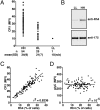
PfRh4 and CR1 directly interact. (A) Immunoprecipitation with anti-PfRh4 monoclonal antibody 10C9 isolates a complex containing both recombinant PfRh4 and sCR1. The anti-PfRh4 monoclonal 10C9 does not immunoprecipitate sCR1 in the absence of recombinant PfRh4. (B) Anti-CR1 monoclonal antibody HB8592 immunoprecipitates sCR1 and recombinant PfRh4, but not another hexaHis-tagged protein (control). (C) An ELISA- based assay measuring the interaction between sCR1 and recombinant PfRh4. Microtiter wells were coated with recombinant PfRh4 or control hexaHis-tagged protein at 0.5 μg/well. sCR1 was added at 0–1 μg/well. Bound CR1 was detected with an anti-CR1 monoclonal HB8592. (D) Use of SPR to measure the dissociation constant (Kd) of sCR1 for PfRh4. (Left) Duplicate sensorgrams for a range of increasing sCR1 concentrations (0.5, 1.0, 2.5, 5.0, and 10 μM, from bottom to top) flowed over a CM5 chip surface with a loading (via amine coupling) of 1,476 response units (RU) of recombinant PfRh4. (Right) Plots of the RUs obtained versus sCR1 concentrations on two different flow cells, coupled with 408 RUs (lower curve) and 1476 RUs (upper curve) of recombinant PfRh4. The dashed vertical line indicates the Kd fitted to both plots simultaneously. (E) Binding of sCR1 to recombinant PfRh4 was inhibited by anti-CR1 monoclonal antibodies. Microtiter plates were coated with saturating concentration of recombinant PfRh4 (5 μg/well). Anti-CR1 monoclonal antibodies and Ig2a mouse isotype at concentrations of 0–1 μg/well were incubated with sCR1 (0.2 μg/well) before being added to the wells. Interaction between sCR1 and PfRh4 was detected using anti-CR1 antibody 6B1 conjugated directly to HRP. 6B1 shares no similar epitopes with the other anti-CR1 antibodies. All ELISA experiments (C and E) were repeated, and similar results were obtained. In C and E, the y axis indicates absorbance measured at 405 nm. Error bars represent the range of duplicate readings. (F) Anti-CR1 antibodies inhibit native PfRh4 erythrocyte binding in an epitope-specific manner. For erythrocyte-binding inhibition assays, the monoclonal antibodies were incubated with erythrocytes at 0.03 mg/mL before the culture supernatants were added. Lane 0 has no antibody added. Immunodetection of parasite proteins with anti-PfRh4 and anti–EBA-175 antibodies after erythrocyte binding is shown.
Fig. 3.
Fig. 3.
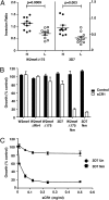
The level of PfRh4 binding correlates with CR1 expression on the erythrocyte surface. (A) Erythrocyte CR1 levels in relation to CR1 genotyping at exon 22 for 80 samples. Each point represents the average of MFI from duplicate readings. H, the high-CR1 allele; L, the low-CR1 allele; n, number of samples; n/a, not applicable. (B) Binding of native PfRh4 to erythrocytes from LL and HH individuals. Immunodetection of parasite proteins with anti-PfRh4 and anti–EBA-175 antibodies after erythrocyte binding is shown. (C) The percentage of recombinant PfRh4-bound erythrocytes (x axis) correlates with the CR1 level on the erythrocyte surface (y axis). Recombinant PfRh4 was added at 0.2 mg/mL to erythrocytes before proceeding with the FACS-based erythrocyte-binding assay, and binding was detected using anti-PfRh4 monoclonal antibody. (D) Percentage of recombinant PfRh4-bound erythrocytes (x axis) does not correlate with glycophorin C expression (y axis). In C and D, r2 is a measure of the goodness of fit of linear regression.
Fig. 4.
Fig. 4.
Sialic acid-independent invasion is reduced in low-CR1 erythrocytes and inhibited in the presence of sCR1. (A) Parasite growth is reduced into neuraminidase-treated low-CR1 erythrocytes. The efficiency of sialic acid-independent invasion in each sample (i.e., the invasion ratio) was calculated as the ratio of the percentage of parasitemia in neuraminidase (Nm)-treated erythrocytes divided by the percentage of parasitemia in untreated erythrocytes using the same strain. These results are shown for invasion into high-CR1 erythrocytes (H, black circles) and low-CR1 erythrocytes (L, white circles) for both W2mefΔ175 (left y axis) and 3D7 (right y axis). The mean ± SEM invasion rates are shown. P values were determined using the unpaired Student t test. (B) The PfRh4 invasion pathway is inhibited in the presence of sCR1. Parasite strains W2mef, W2mefΔRh4, W2mefΔ175, and 3D7 were tested in growth assays into untreated or Nm-treated erythrocytes in the presence of 0.5 mg/mL of sCR1 (white bars) or control protein (black bars). (C) Inhibition of parasite growth by sCR1 is concentration-dependent. Invasion of 3D7 into untreated (un) or Nm-treated erythrocytes was measured in the presence of sCR1at the stated 0–0.5 mg/mL final concentration. In B and C, growth is measured as the percent of noninhibitory PBS control, and error bars represent the SEM from two separate experiments (in triplicate).

References

    1. Cowman AF, Crabb BS. Invasion of red blood cells by malaria parasites. Cell. 2006;124:755–766. - PubMed
    1. Peterson DS, Wellems TE. EBL-1, a putative erythrocyte-binding protein of Plasmodium falciparum, maps within a favored linkage group in two genetic crosses. Mol Biochem Parasitol. 2000;105:105–113. - PubMed
    1. Adams JH, et al. A family of erythrocyte-binding proteins of malaria parasites. Proc Natl Acad Sci USA. 1992;89:7085–7089. - PMC - PubMed
    1. Rayner JC, Galinski MR, Ingravallo P, Barnwell JW. Two Plasmodium falciparum genes express merozoite proteins that are related to Plasmodium vivax and Plasmodium yoelii adhesive proteins involved in host cell selection and invasion. Proc Natl Acad Sci USA. 2000;97:9648–9653. - PMC - PubMed
    1. Rayner JC, Vargas-Serrato E, Huber CS, Galinski MR, Barnwell JW. A Plasmodium falciparum homologue of Plasmodium vivax reticulocyte binding protein (PvRBP1) defines a trypsin-resistant erythrocyte invasion pathway. J Exp Med. 2001;194:1571–1581. - PMC - PubMed

Publication types

MeSH terms

LinkOut - more resources