. 2010 Oct 5;107(40):17188-93.
doi: 10.1073/pnas.1012348107. Epub 2010 Sep 20.

Mutations of cellulose synthase (CESA1) phosphorylation sites modulate anisotropic cell expansion and bidirectional mobility of cellulose synthase

Affiliations

Mutations of cellulose synthase (CESA1) phosphorylation sites modulate anisotropic cell expansion and bidirectional mobility of cellulose synthase

Shaolin Chen et al. Proc Natl Acad Sci U S A. .

Abstract

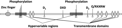
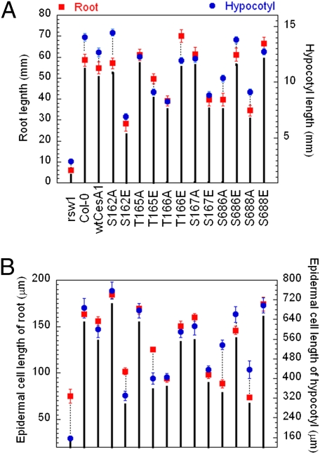
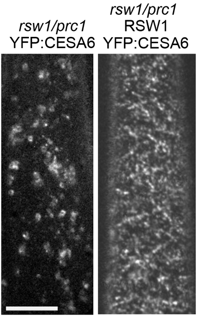
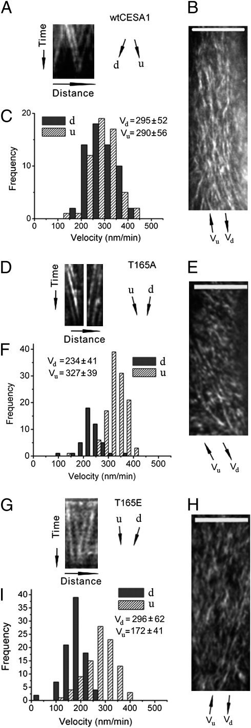
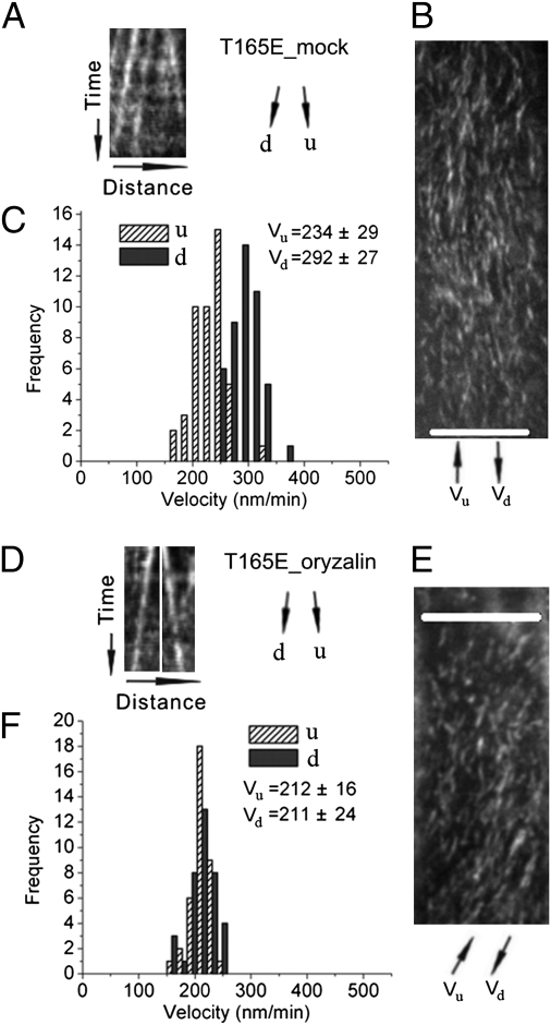
The CESA1 component of cellulose synthase is phosphorylated at sites clustered in two hypervariable regions of the protein. Mutations of the phosphorylated residues to Ala (A) or Glu (E) alter anisotropic cell expansion and cellulose synthesis in rapidly expanding roots and hypocotyls. Expression of T166E, S686E, or S688E mutants of CESA1 fully rescued the temperature sensitive cesA1-1 allele (rsw1) at a restrictive temperature whereas mutations to A at these positions caused defects in anisotropic cell expansion. However, mutations to E at residues surrounding T166 (i.e., S162, T165, and S167) caused opposite effects. Live-cell imaging of fluorescently labeled CESA showed close correlations between tissue or cell morphology and patterns of bidirectional motility of CESA complexes in the plasma membrane. In the WT, CESA complexes moved at similar velocities in both directions along microtubule tracks. By contrast, the rate of movement of CESA particles was directionally asymmetric in mutant lines that exhibited abnormal tissue or cell expansion, and the asymmetry was removed upon depolymerizing microtubules with oryzalin. This suggests that phosphorylation of CESA differentially affects a polar interaction with microtubules that may regulate the length or quantity of a subset of cellulose microfibrils and that this, in turn, alters microfibril structure in the primary cell wall resulting in or contributing to the observed defect in anisotropic cell expansion.


Conflict of interest statement

The authors declare no conflict of interest.

Figures

Fig. 1.
Fig. 1.
Illustration of the structure of CESA1 (1081 aa). Indicated phosphorylation sites include S162, T165, T166, S167, S686, and S688.
Fig. 2.
Fig. 2.
Lengths of primary roots and hypocotyls (A) and their epidermal cells at the mature zone (B) of Col-0, rsw1, and rsw1 transgenic lines containing WT CESA1 cDNA or cesA1P mutants. For root measurements, seedlings were grown in continuous light (200 μmol/m2/s) on vertical 0.5× MS agar plates at 30 °C for 7 d. For hypocotyl measurements, seedlings were grown on vertical 0.5× MS agar plates at 30 °C for 5 d in darkness. Mean values are shown with SEs (≥50 measurements).
Fig. 3.
Fig. 3.
Effect of temperature on the accumulation of YFP::CESA6 in Golgi and plasma membrane in an rsw1 prc1 background. Etiolated seedlings were grown at restrictive temperature (30 °C) for 2 d, then used to visualize CESA complexes at 29 °C. YFP::CESA6 localization in etiolated hypocotyl cells of rsw1/prc1-1 seedlings expressing YFP::CESA6 (Left) or both YFP::CESA6 and WT CESA1 (Right). Both transgenic lines are heterozygous for YFP::CESA6. (Scale bar: 10 μm.)
Fig. 4.
Fig. 4.
Kymograph analysis of bidirectional movement of CESA complexes in the WT, T165A, and T165E transgenic line. The data for the other mutants is presented in Fig. S4. Time-lapse confocal images of YFP::CESA6 in hypocotyl cells of etiolated seedlings grown at restrictive temperature for 2 d were used to measure CESA particle velocity at 29 °C. To analyze bidirectional movement of CESA particles, we defined the average axis of CESA particle movement in a given image series of a cell as the major axis and classified CESA particles as moving in upward (u) or downward (d) direction along the major axis, as illustrated in Fig. S4 A and B. (A, D, and G) Representative kymograph displays effects of mutations on bidirectional particle translocations, derived from kymograph analysis of CESA particles moving along common linear tracks. (B, E, and H) Average time projections of 61 frames representing 5 min. (C, F, and I) Histograms of particle velocities calculated from kymograph analysis of 100 to 200 particles from three to six cells of a single seedling. Similar results were obtained from different cells in different seedlings. (Scale bars: 10 μm in B, E, and H.)
Fig. 5.
Fig. 5.
Effects of oryzalin on bidirectional movement of CESA complexes in the T165E line. Time-lapse confocal images of YFP::CESA6 in hypocotyl cells of 2-d-old etiolated plants grown at 30 °C were used to measure CESA particle velocity. u and d represent two opposite directions of particle movement relative to the major axis (Fig. S4 A and B). (AC) Control treatment with 0.02% methanol or DMSO at 30 °C for 2 to 3 h. (DF) Treatment with 20 μM oryzalin at 30 °C for 2 to 3 h. (A and D) Representative kymograph displays effects of oryzalin treatment on bidirectional particle translocation. (B and E) Average time projections of 61 frames representing 5 min. (C and F) Histograms of particle velocities calculated from kymograph analysis of 100 to 200 particles from three to six cells of a single seedling. (Scale bars: 10 μm in B and E.)

References

    1. Somerville C. Cellulose synthesis in higher plants. Annu Rev Cell Dev Biol. 2006;22:53–78. - PubMed
    1. Arioli T, et al. Molecular analysis of cellulose biosynthesis in Arabidopsis. Science. 1998;279:717–720. - PubMed
    1. Heim DR, Skomp JR, Tschabold EE, Larrinua IM. Isoxaben inhibits the synthesis of acid insoluble cell-wall materials in Arabidopsis thaliana. Plant Physiol. 1990;93:695–700. - PMC - PubMed
    1. Roudier F, et al. COBRA, an Arabidopsis extracellular glycosyl-phosphatidyl inositol-anchored protein, specifically controls highly anisotropic expansion through its involvement in cellulose microfibril orientation. Plant Cell. 2005;17:1749–1763. - PMC - PubMed
    1. Saxena IM, Brown RM., Jr Cellulose biosynthesis: Current views and evolving concepts. Ann Bot (Lond) 2005;96:9–21. - PMC - PubMed

Publication types

MeSH terms