. 2010 Oct 5;107(40):17182-7.
doi: 10.1073/pnas.1012556107. Epub 2010 Sep 20.

Lateral opening of a translocon upon entry of protein suggests the mechanism of insertion into membranes

Affiliations

Lateral opening of a translocon upon entry of protein suggests the mechanism of insertion into membranes

Pascal F Egea et al. Proc Natl Acad Sci U S A. .

Abstract

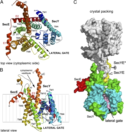
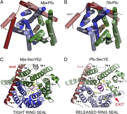
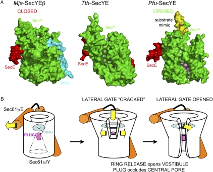
The structure of the protein-translocating channel SecYEβ from Pyrococcus furiosus at 3.1-Å resolution suggests a mechanism for chaperoning transmembrane regions of a protein substrate during its lateral delivery into the lipid bilayer. Cytoplasmic segments of SecY orient the C-terminal α-helical region of another molecule, suggesting a general binding mode and a promiscuous guiding surface capable of accommodating diverse nascent chains at the exit of the ribosomal tunnel. To accommodate this putative nascent chain mimic, the cytoplasmic vestibule widens, and a lateral exit portal is opened throughout its entire length for partition of transmembrane helical segments to the lipid bilayer. In this primed channel, the central plug still occludes the pore while the lateral gate is opened, enabling topological arbitration during early protein insertion. In vivo, a 15 amino acid truncation of the cytoplasmic C-terminal helix of SecY fails to rescue a secY-deficient strain, supporting the essential role of this helix as suggested from the structure.


Conflict of interest statement

The authors declare no conflict of interest.

Figures

Fig. 1.
Fig. 1.
Overview of the structure of Pfu-SecYE and the crystal packing. SecY is colored using a rainbow pattern. The plug (α3) and C-terminal (αC) helices are labeled. (A) Top view showing the cytoplasmic vestibule and the pore occluded by the plug. The two halves constituting SecY are delineated with an arrow indicating the lateral opening. (B) View showing the lateral gate opening between transmembrane helices TM2 and TM3 (in the N-terminal one-half) and TM7 and TM8 (in the C-terminal one-half). The yellow line delineates the lateral gate on the SecY subunit. (C) Surface representation of two complexes as they pack in the crystal. A SecYE complex interacts through the insertion of its C-terminal αC-helix in the cytoplasmic vestibule of a crystallographically related complex (SecYE* in gray). The plug and αC-helices are colored in pink and yellow, respectively. TMs delineating the opened lateral gate are colored in cyan.
Fig. 2.
Fig. 2.
Conformational changes observed in the translocons from Pyrococcus, Thermus, and Methanococcus. Superposition of the Mja and Pfu channels (A) and the Tth and Pfu channels (B). The top view from the cytoplasmic side emphasizes the rearrangement of the N terminus of SecE and the rigid body motions of SecY TMs. The plug helix remains in place. TMs 2, 3, 7, and 8 delineate the lateral gate. The SecE subunit is colored in red. The arrows show movements from Mja or Tth to Pfu. The hydrophobic seal provided by the ring region is compromised on lateral gate opening. Cytoplasmic view along the channel pore showing the rearrangement of the hydrophobic ring that seals the cytoplasmic vestibule of the channel above the plug region. (C) Mja structure with a closed ring. (D) Pfu structure with an open ring. The N- and C-terminal halves of SecY are colored in green and blue, respectively. TMs and ring residues are labeled. The plug helix is colored in pink. Pfu is in light colors, and Mja or Tth is in dark colors.
Fig. 3.
Fig. 3.
The lateral gate of the channel of the Pfu-SecYE channel is entirely opened while its plug remains in place, still occluding the central conduit. (A) Surface representations of the translocons from Mja, Tth, and Pfu showing the different states of the lateral gate. The SecY, SecE, and Secβ subunits are colored in green, red, and cyan, respectively. The αC-helix contributed by a symmetry-related SecYE complex is shown as a yellow surface to emphasize its insertion into the cytoplasmic vestibule. In Tth and Pfu, the so-called hydrophobic crack observed in Tth-SecYE (8) and the complete lateral gate opening in Pfu-SecYE are shown as a dotted white line. The plug helix is colored in pink and can only be clearly seen in the Pfu structure because of the extensive lateral gate. (B) Schematics of the three conformations shown in A.
Fig. 4.
Fig. 4.
In vivo rescue of the secY-deficient bacterial strain by the archaeal SecY requires its C-terminal cytoplasmic helix. Complementation of the secY24 ts mutant E. coli strain with vectors expressing no gene (empty), the E. coli-SecY or Pfu-SecY gene clusters in the case of complete truncation (A), or point mutations (B) in the cytoplasmic C terminus of Pfu-SecY. The growth patterns at permissive and nonpermissive temperatures are shown. Pfu-Y and Ec-Y segments indicate the presence of wild-type full-length proteins; Δ segments indicate the presence of C-terminal deletion mutants in which 15 residues (454–468) are deleted from Pfu-SecY or 20 residues (424–443) are deleted from E. coli-SecY. Point mutants in the full-length Pfu protein are labeled E457A, F459A, R463A, K464A, R463A/K464A, and F459P/A461P. (C) Model of the ribosome/translocon interaction based on our structure aligned and the Sec61-translocon cryo-EM structure (12). At the ribosomal tunnel exit site, the C-terminal helix of SecY (red) packs against ribosomal RNAs (yellow) and proteins (blue). Δ indicates the position of the deletion. The sequence of Pfu-SecY is shown; the area shaded in yellow indicates the C-terminal truncation. Membrane boundaries are depicted as brown lines.

References

    1. White SH, von Heijne G. The machinery of membrane protein assembly. Curr Opin Struct Biol. 2004;14:397–404. - PubMed
    1. Brundage L, Hendrick JP, Schiebel E, Driessen AJ, Wickner W. The purified E. coli integral membrane protein SecY/E is sufficient for reconstitution of SecA-dependent precursor protein translocation. Cell. 1990;62:649–657. - PubMed
    1. Meyer TH, et al. The bacterial SecY/E translocation complex forms channel-like structures similar to those of the eukaryotic Sec61p complex. J Mol Biol. 1999;285:1789–1800. - PubMed
    1. Egea PF, Stroud RM, Walter P. Targeting proteins to membranes: Structure of the signal recognition particle. Curr Opin Struct Biol. 2005;15:213–220. - PubMed
    1. Rusch SL, Kendall DA. Interactions that drive Sec-dependent bacterial protein transport. Biochemistry. 2007;46:9665–9673. - PMC - PubMed

Publication types

Associated data