. 2010 Oct 5;107(40):17362-7.
doi: 10.1073/pnas.1012568107. Epub 2010 Sep 20.

Soluble amyloid precursor protein (APP) regulates transthyretin and Klotho gene expression without rescuing the essential function of APP

Affiliations

Soluble amyloid precursor protein (APP) regulates transthyretin and Klotho gene expression without rescuing the essential function of APP

Hongmei Li et al. Proc Natl Acad Sci U S A. .

Erratum in

  • Proc Natl Acad Sci U S A. 2013 Aug 6;110(32):13228

Abstract

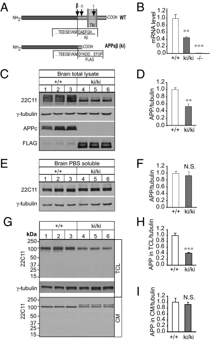
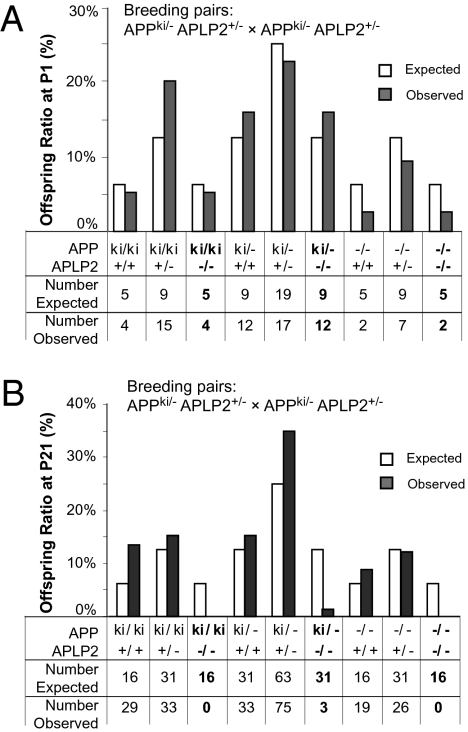
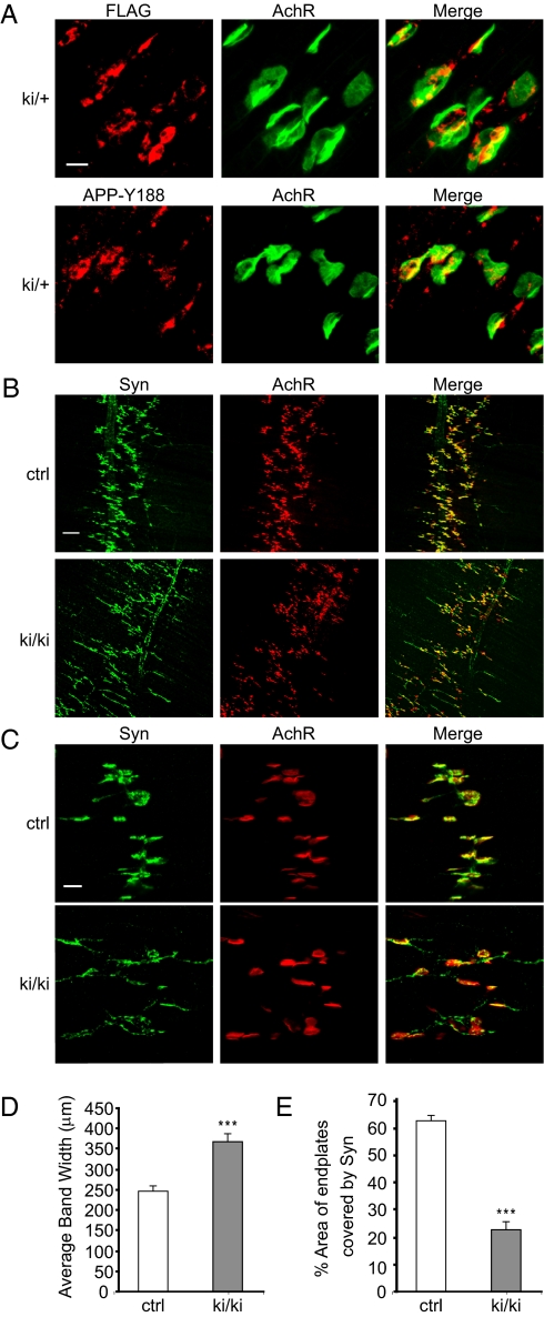
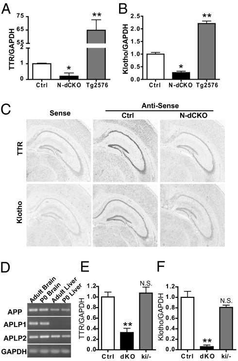
Amyloidogenic processing of the amyloid precursor protein (APP) generates a large secreted ectodomain fragment (APPsβ), β-amyloid (Aβ) peptides, and an APP intracellular domain (AICD). Whereas Aβ is viewed as critical for Alzheimer's disease pathogenesis, the role of other APP processing products remains enigmatic. Of interest, the AICD has been implicated in transcriptional regulation, and N-terminal cleavage of APPsβ has been suggested to produce an active fragment that may mediate axonal pruning and neuronal cell death. We previously reported that mice deficient in APP and APP-like protein 2 (APLP2) exhibit early postnatal lethality and neuromuscular synapse defects, whereas mice with neuronal conditional deletion of APP and APLP2 are viable. Using transcriptional profiling, we now identify transthyretin (TTR) and Klotho as APP/APLP2-dependent genes whose expression is decreased in loss-of-function states but increased in gain-of-function states. Significantly, by creating an APP knockin allele that expresses only APPsβ protein, we demonstrate that APPsβ is not normally cleaved in vivo and is fully capable of mediating the APP-dependent regulation of TTR and Klotho gene expression. Despite being an active regulator of gene expression, APPsβ did not rescue the lethality and neuromuscular synapse defects of APP and APLP2 double-KO animals. Our studies identify TTR and Klotho as physiological targets of APP that are regulated by soluble APPsβ independent of developmental APP functions. This unexpected APP-mediated signaling pathway may play an important role in maintaining TTR and Klotho levels and their respective functions in Aβ sequestration and aging.


Conflict of interest statement

The authors declare no conflict of interest.

Figures

Fig. 1.
Fig. 1.
Generation and biochemical characterization of APPsβ ki mice. (A) Schematic representation of WT and APPsβ ki alleles. Cleavage sites for α-, β-, and γ-secretase are indicated by α, β, and γ, respectively. In the ki allele, a FLAG sequence followed by a stop codon was inserted immediately downstream of the β-secretase site. TM, transmembrane region. (B) qRT-PCR of APP mRNA in 2-mo-old homozygous APPsβ ki/ki and APP KO (−/−) mouse brains relative to WT controls (+/+), showing an ∼50% reduction of APP mRNA in ki/ki samples and a negligible amount in APP−/− samples as compared with the WT controls. (C) Western blot analysis of APP expression in total brain lysate from 2-mo-old ki/ki mice and their WT (+/+) littermates using the 22C11, APPc, and anti-FLAG antibodies. A γ-tubulin blot was used as a loading control. (D) Quantification of the relative ratio of 22C11/γ-tubulin blots. (E) Western blot analysis of PBS-extractable soluble APP in +/+ and ki/ki brains using the 22C11 antibody. A γ-tubulin blot was used as a protein loading control. (F) Quantification of the Western blots in E. (G) Representative Western blots of APP in TCL and CM from cultured WT (+/+) and homozygous APPsβ (ki/ki) primary neurons using the 22C11 antibody. A γ-tubulin blot was used as a loading control. (H) Quantification of the ratio of APP/tubulin in TCL of +/+ and ki/ki cultures. (I) Quantification of the ratio of APP/tubulin in CM of +/+ and ki/ki cultures. Note the absence of any cleavage products in the TCL or CM. The ratio of APP/tubulin in +/+ samples was normalized to 1 in all quantifications. **P < 0.01; ***P < 0.001. N.S., nonsignificant (P > 0.05, t test).
Fig. 2.
Fig. 2.
Survival analysis of APPsβ ki mice on APLP2 null background. (A) Analysis of genotypes of 75 offspring collected at P1 derived from crosses of APPki/−APLP2+/− male and female mice. All genotypes were recovered at close to a Mendelian ratio (8 df, χ2 = 10.65, P > 0.1). (B) Analysis of genotypes of 218 offspring collected at P21 derived from the same breeding as in A. The number of APPki/kiAPLP2−/−, APPki/−APLP2−/−, or APP−/−APLP2−/− animals observed was much lower than expected (highlighted in bold) (8 df, χ2 = 79.6, P < 0.001).
Fig. 3.
Fig. 3.
Analysis of neuromuscular synapse phenotypes of APPsβ ki mice. (A) Immunofluorescence staining of P0 sternomastoid muscle sections of heterozygous APPsβ ki animals (ki/+) using an anti-FLAG antibody and the anti-APP antibody Y188. Staining with α-bungarotoxin was used to mark the postsynaptic AchRs. (Merge) Overlay of APP and AchR images. (B) Whole-mount staining of P0 diaphragm muscles of APPki/kiAPLP2−/− mutants (ki/ki) and littermate APP+/+APLP2−/− controls (ctrl) with an anti-synaptophysin (Syn) antibody and α-bungarotoxin (AchR) showing diffused pre- and postsynaptic distribution in the ki/ki mutant. (Merge) Overlay of Syn and AchR images. (C) Higher magnification images of synapse structures showing axonal staining of Syn and poorly covered end plates by Syn in the ki/ki mutant. (D) Quantification of the average bandwidth of AchR-positive end plates. (E) Quantification of the percentage of AchR-positive end plates covered by Syn (average ± SEM of 20 end plates per genotype). ***P < 0.001 (Student's t test). (Scale bar: A and C, 20 μM; B, 100 μM.)
Fig. 4.
Fig. 4.
Analysis of TTR and Klotho expression. Relative qRT-PCR analysis of TTR (A) and Klotho (B) mRNA levels from the hippocampi of 2-mo-old control (Ctrl), N-dCKO, and Tg2576 APP transgenic mice (n = 3). (C) Representative in situ hybridization images of TTR and Klotho from littermate APLP2−/− control (Ctrl) and N-dCKO brain sections (sense, hybridization with a sense probe as a negative control; antisense, hybridization using corresponding antisense probes). (D) RT-PCR analysis of APP, APLP1, and APLP2 expression in adult and P0 brain and liver. GAPDH was used as an amplification control. Relative qRT-PCR analysis of TTR (E) and Klotho (F) mRNA levels in P0 livers from control APLP2 null (Ctrl), APP, and APLP2 dKO and APPsβ ki mice on APLP2 null background (ki/−). Data are the mean ± SEM of two independent experiments, each with three samples per genotype. *P < 0.05; **P < 0.01. (N.S., nonsignificant, Student's t test).

References

    1. Zheng H, Koo EH. The amyloid precursor protein: Beyond amyloid. Mol Neurodegener. 2006;1:5. - PMC - PubMed
    1. Turner PR, O'Connor K, Tate WP, Abraham WC. Roles of amyloid precursor protein and its fragments in regulating neural activity, plasticity and memory. Prog Neurobiol. 2003;70:1–32. - PubMed
    1. Nikolaev A, McLaughlin T, O'Leary DDM, Tessier-Lavigne M. APP binds DR6 to trigger axon pruning and neuron death via distinct caspases. Nature. 2009;457:981–989. - PMC - PubMed
    1. Cao X, Südhof TC. A transcriptionally active complex of APP with Fe65 and histone acetyltransferase Tip60. Science. 2001;293:115–120. - PubMed
    1. Cao X, Südhof TC. Dissection of amyloid-beta precursor protein-dependent transcriptional transactivation. J Biol Chem. 2004;279:24601–24611. - PubMed

Publication types

MeSH terms