. 2010 Oct 5;107(40):17211-6.
doi: 10.1073/pnas.1007032107. Epub 2010 Sep 20.

Sexual dimorphism of gonadal structure and gene expression in germ cell-deficient loach, a teleost fish

Affiliations

Sexual dimorphism of gonadal structure and gene expression in germ cell-deficient loach, a teleost fish

Takafumi Fujimoto et al. Proc Natl Acad Sci U S A. .

Abstract

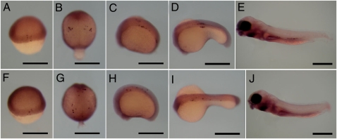
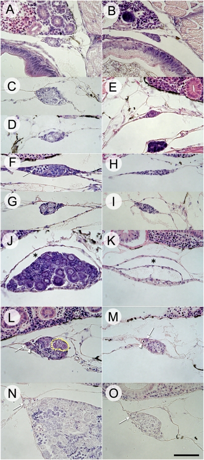
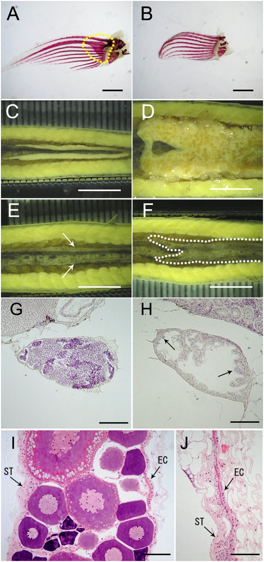
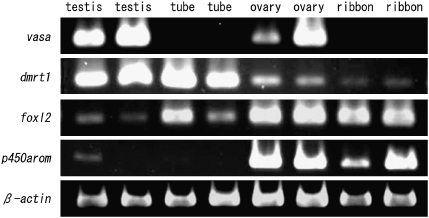
Germ cell-deficient fish usually develop as phenotypic males. Thus, the presence of germ cells is generally considered to be essential for female gonadal differentiation or the maintenance of ovarian structure. However, little is known of the role of germ cells in the determination of the sexual fate of gonadal somatic cells. We have established an inducible germ cell deficiency system in the loach (Misgurnus anguillicaudatus, Cypriniformes: Cobitidae), a small freshwater fish, using knockdown of the dead end gene with a morpholino antisense oligonucleotide. Interestingly, loach lacking germ cells could develop as either phenotypic males or females, as characterized morphologically by the presence or absence of bony plates in the pectoral fins, respectively. The phenotypic males and females had testicular and ovarian structures, respectively, but lacked germ cells. Gene expression patterns in these male and female germ cell-deficient gonads were essentially the same as those in gonads of normal fish. Our observations indicate that sexually dimorphic gonads can develop in germ cell-deficient loach. In contrast to the situation in other model fish species, the gonadal somatic cells in phenotypic females autonomously differentiated into ovarian tissues and also played a role in the maintenance of gonadal structure. On the basis of our observations, we propose two possible models to explain the role of germ cells in sex determination in fish.


Conflict of interest statement

The authors declare no conflict of interest.

Figures

Fig. 1.
Fig. 1.
Migrating primordial germ cells (PGCs) were identified by vasa expression using whole mount in situ hybridization of wild-type and dnd-MO injected (2,000–4,000 pg/embryo) embryos at the embryonic shield to larval stages. (AE and FJ) PGCs of wild-type and dnd morphants, respectively. (A and F) Embryonic shield stage. (B and G) Two- to four-somite stage. (C and H) Twenty-somite stage. (D and I) Thirty-somite stage. (E and J) Larvae at 5 d postfertilization. (Scale bars, 500 μm.)
Fig. 2.
Fig. 2.
Sex differentiation in wild-type fish and dnd morphants during gonadal development. (A) Wild-type gonad at 33 d posthatch (dph). (B) dnd morphant gonad at 33 dph. (C) Wild-type gonad with proliferating germ cells within a cyst at 55 dph. (D) Wild-type gonad with only a few germ cells and no cyst formation at 55 dph. (E) dnd morphant gonad at 55 dph. (F) Laterally elongated gonad with proliferating germ cells within a cyst in a wild-type gonad at 70 dph. Arrow indicates germ cells at the zygotene stage of meiosis. (G) Club-shaped gonad with a few germ cells in a wild-type gonad at 70 dph. (H) Laterally elongated gonad in dnd morphant at 70 dph. (I) Club-shaped gonad in dnd morphant at 70 dph. (J) Ovary with oocytes in wild type at 97 dph. Asterisk indicates ovarian lumen. (K) Ribbon-like gonad of dnd morphant at 120 dph. Asterisk indicates ovarian lumen. (L) Testis with undifferentiated germ cells in a wild-type gonad at 97 dph. Arrow and arrowhead indicate sperm duct anlage and blood vessel, respectively. Dotted line indicates seminal lobules. (M) Tube-like gonad of dnd morphant at 120 dph. Arrow and arrowhead indicate sperm duct anlage and blood vessel, respectively. (N) Testis with germ cells undergoing spermatogenesis in a wild-type gonad at 150 dph. Arrow indicates sperm duct. (O) Tube-like gonad of dnd morphant at 150 dph. Arrow indicates sperm duct. (Scale bar, 50 μm.)
Fig. 3.
Fig. 3.
Sexual dimorphism in 2-y-old wild-type fish and dnd morphants. (A) Pectoral fin of male with bony plate (framed by dotted line). (B) Pectoral fin of females without bony plate. (C) Testes in wild-type male. (D) Ovary in wild-type female. (E) Tube-like gonad (indicated by arrows) in a phenotypically male dnd morphant. (F) Ribbon-like gonad (indicated by dotted line) in a phenotypically female dnd morphant. (G) Testis of wild-type male. (H) Tube-like gonad of male dnd morphant. (I) Ovary of wild-type female. (J) Ribbon-like gonad of female dnd morphant. EC, epithelial cells; ST, stromal tissue. [Scale bars, 1 mm (A and B), 5 mm (CF), and 50 μm (GJ).]
Fig. 4.
Fig. 4.
Patterns of expression of vasa, dmrt1, foxl2, p450arom, and β-actin in gonads of wild-type fish and dnd morphants. Two replicate RT-PCR analyses were performed using independent samples from each gonadal type.

References

    1. Devlin RH, Nagahama Y. Sex determination and sex differentiation in fish: an overview of genetic, physiological, and environmental influences. Aquaculture. 2002;208:191–364.
    1. Baroiller JF, D'Cotta H, Saillant E. Environmental effects on fish sex determination and differentiation. Sex Dev. 2009;3:118–135. - PubMed
    1. Takahashi H. Juvenile hermaphroditism in the zebrafish, Brachydanio rerio. Bull Fac Fish Hokkaido Univ. 1977;28:57–65.
    1. Matsuda M, et al. DMY is a Y-specific DM-domain gene required for male development in the medaka fish. Nature. 2002;417:559–563. - PubMed
    1. Matsuda M. Sex determination in the teleost medaka, Oryzias latipes. Annu Rev Genet. 2005;39:293–307. - PubMed

Publication types

Substances

LinkOut - more resources