. 2010 Oct 5;107(40):17356-61.
doi: 10.1073/pnas.1008306107. Epub 2010 Sep 20.

Oscillatory phase coupling coordinates anatomically dispersed functional cell assemblies

Affiliations

Oscillatory phase coupling coordinates anatomically dispersed functional cell assemblies

Ryan T Canolty et al. Proc Natl Acad Sci U S A. .

Abstract

Hebb proposed that neuronal cell assemblies are critical for effective perception, cognition, and action. However, evidence for brain mechanisms that coordinate multiple coactive assemblies remains lacking. Neuronal oscillations have been suggested as one possible mechanism for cell assembly coordination. Prior studies have shown that spike timing depends upon local field potential (LFP) phase proximal to the cell body, but few studies have examined the dependence of spiking on distal LFP phases in other brain areas far from the neuron or the influence of LFP-LFP phase coupling between distal areas on spiking. We investigated these interactions by recording LFPs and single-unit activity using multiple microelectrode arrays in several brain areas and then used a unique probabilistic multivariate phase distribution to model the dependence of spike timing on the full pattern of proximal LFP phases, distal LFP phases, and LFP-LFP phase coupling between electrodes. Here we show that spiking activity in single neurons and neuronal ensembles depends on dynamic patterns of oscillatory phase coupling between multiple brain areas, in addition to the effects of proximal LFP phase. Neurons that prefer similar patterns of phase coupling exhibit similar changes in spike rates, whereas neurons with different preferences show divergent responses, providing a basic mechanism to bind different neurons together into coordinated cell assemblies. Surprisingly, phase-coupling-based rate correlations are independent of interneuron distance. Phase-coupling preferences correlate with behavior and neural function and remain stable over multiple days. These findings suggest that neuronal oscillations enable selective and dynamic control of distributed functional cell assemblies.


Conflict of interest statement

The authors declare no conflict of interest.

Figures

Fig. 1.
Fig. 1.
Patterns of oscillatory phase coupling across multiple brain areas coordinate anatomically dispersed neuronal cell assemblies (schematic). (A–D) Hypothesis 1: Spike timing in single neurons depends on frequency-specific oscillatory phase coupling across multiple brain areas. (A) Spiking in one area may depend on population activity (local field potentials, LFPs) occurring in multiple areas. (B) Many neurons are sensitive to oscillatory LFP activity occurring in particular frequency bands; filtering all LFPs at this frequency and extracting phases can reveal patterns of phase coupling between LFP channels. (C) The strength of LFP–LFP phase coupling is different for spike times compared with randomly selected times and defines a neuron's preferred pattern of LFP–LFP phase coupling, similar to a receptive field. That is, when LFP activity matches the neuron's preferred pattern of LFP–LFP phase coupling, the cell spikes more often. (D) Given novel LFP phases as input, the model generates a predicted coupling-based spike rate output, which can then be compared with the measured spike rate. (EG) Hypothesis 2: Large-scale patterns of phase coupling synchronize anatomically dispersed neuronal ensembles. (E) The procedure described above can be applied to multiple simultaneously recorded neurons. (F) Cells that prefer similar LFP–LFP phase-coupling patterns exhibit similar coupling-based rates. (G) Shared variability in coupling-based rates is compactly described by a single phase coupling network that defines a cell assembly. That is, it is possible to identify large-scale patterns of LFP–LFP phase coupling (G) that explain a significant fraction of the variation in spike rates for a large ensemble of neurons distributed across multiple brain areas. (H–J) Hypothesis 3: Differential sensitivity to distinct brain rhythms or coupling patterns permits selective control of multiple coactive assemblies. (H) Multiple functional ensembles, each spanning several brain areas, overlap in space. (I) Interference between ensembles is minimized when each assembly responds to a different frequency (assemblies A and C) or distinct phase-coupling pattern (assemblies A and B). (J) Frequency and pattern selectivity permits dynamic, independent coordination of multiple coactive ensembles.
Fig. 2.
Fig. 2.
Spike timing in single neurons depends on oscillatory phase coupling between multiple brain areas. (A) Example of a neuron where the probability of spiking depends on frequency-specific LFP phase in multiple areas. The neuron is located in right primary motor cortex (M1). Colored traces represent different LFPs recorded from left M1 (blue), right M1 (green), or left dorsal premotor area (PMd, red). The strong high β (25–40 Hz)-modulation shown here is typical of M1-PMd neurons. (B–G) Estimates of spike/LFP interactions depend on the method used and some commonly used techniques may generate misleading results. Examining all LFP signals at once results in differing estimates of phase coupling strength compared with examining pairs of LFP signals separately, as shown by differences between empirical (black) and isolated (red) probability density functions (main text and SI Methods; also Figs. S4 and S5). (H) Preferred phase coupling network representing 48 LFPs from three brain areas for the M1 neuron shown in A. Nodes represent LFP phase variables; links represent the strength of LFP–LFP phase coupling, from weak (light lines) to strong (dark lines). Node size is proportional to the sum of link connection weights entering the node. Strong cross-area coupling remains after conditioning on proximal/distal phases and within-area phase coupling. This preferred pattern of phase coupling acts like an internal, LFP-based receptive field; when the instantaneous pattern of phase coupling between electrodes is close to the preferred coupling pattern, the cell spikes more often. (I) The coupling-based spike rate (generated from the preferred LFP–LFP phase coupling pattern learned from training data and instantaneous LFP phases from test data) predicts the measured spike rate (calculated using spike times from test data). (J) The relationship between predicted and measured spike rates is stable over multiple days.
Fig. 3.
Fig. 3.
Large-scale patterns of phase coupling synchronize anatomically dispersed neuronal ensembles. Neurons that prefer similar LFP–LFP phase coupling patterns show correlations between coupling-based spike rates, independent of distance. (A and B) Two neurons from left and right M1 with correlated coupling-based spike rates. (C and D) Two neurons recorded from one microelectrode exhibit a weak coupling-based spike rate correlation despite close spatial proximity. (E) Within a cortical area, coupling-based spike rate correlations do not depend on interneuron distance (5,716 pairs, n.s.). (F) In contrast, motor cortical neurons with similar direction tuning measured during a center-out BMI movement task (28) exhibit coupling-based spike rate correlations (9,413 pairs, P < 0.001), suggesting that coupling-based spike rate correlations depend on neural function but not spatial location. (G) Shared variability in coupling-based rates is concentrated by independent components analysis (ICA), with a small set of components (red) accounting for most of the predictive value of coupling-based rates (see text). (H) Correlation matrix of ICA-denoised coupling-based rates, sorted to identify clusters of neurons with similar activity; e.g., neurons 1–10 form a spatially distributed ensemble with correlated coupling-based rates, have a low correlation with the activity of neurons 61–70, and are anticorrelated with neurons 121–130. The LFP–LFP phase coupling patterns associated with these ICA components explain a portion of the internally generated spike rate variations across an ensemble of anatomically distributed cells and may therefore bind these cells into a functional assembly via Hebbian synaptic modification.
Fig. 4.
Fig. 4.
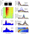
Phase coupling networks exhibit behavior-related changes and may selectively respond to different frequency bands. (A) Monkeys engaged in a brain–machine interface (BMI) task, using some cells (BMI neurons) to drive an on-screen cursor (28). The percentage of neurons exhibiting significant coupling-based rate modulation was the same for BMI and non-BMI groups. (B) Predicted spike rates generated by assembly-specific coupling patterns (SI Methods) show event-related changes. Time is relative to GO cue onset; the vertical axis shows different independent components sorted by activity level 100 ms after cue onset. (C) Peristimulus time histogram (PSTH, blue) for a PMd neuron shows an activity peak at cue onset. The cue-locked average of one coupling-based rate (red) shows event-related changes that correlate with PSTH activity. (D) As in C, for a M1 neuron and different coupling-based rate. Both PSTH (blue) and coupling-based rate (red) peak ∼100 ms after cue. (E–H) Different neurons are sensitive to distinct frequencies. Plots show neuronal sensitivity to LFP phase versus frequency (compare with Fig. 2A). Colored traces represent LFPs from different areas: right (red) and left (yellow) dorsolateral prefrontal cortex, right (blue) and left (green) orbitofrontal cortex, and left cingulate sulcus (black). (I) Eight hundred thirteen neurons from four subjects sorted by preferred frequency (black dots). Horizontal lines show normalized modulation strength from low (blue) to high (red) versus frequency; the broad range of preferred frequencies may enable multiple ensembles to operate with minimal interference.

References

    1. Kandel E. Principles of Neural Science. 4th Ed. New York: McGraw–Hill; 2000.
    1. Destexhe A, Rudolph M, Paré D. The high-conductance state of neocortical neurons in vivo. Nat Rev Neurosci. 2003;4:739–751. - PubMed
    1. Shepherd GM, Stepanyants A, Bureau I, Chklovskii D, Svoboda K. Geometric and functional organization of cortical circuits. Nat Neurosci. 2005;8:782–790. - PubMed
    1. Douglas RJ, Martin KAC. Neuronal circuits of the neocortex. Annu Rev Neurosci. 2004;27:419–451. - PubMed
    1. Hebb DO. The Organization of Behavior. New York: Wiley; 1949.

Publication types

LinkOut - more resources