. 2010 Oct 5;107(40):17262-7.
doi: 10.1073/pnas.1009304107. Epub 2010 Sep 20.

Evidence for the prepattern/cooption model of vertebrate jaw evolution

Affiliations

Evidence for the prepattern/cooption model of vertebrate jaw evolution

Robert Cerny et al. Proc Natl Acad Sci U S A. .

Abstract

The appearance of jaws was a turning point in vertebrate evolution because it allowed primitive vertebrates to capture and process large, motile prey. The vertebrate jaw consists of separate dorsal and ventral skeletal elements connected by a joint. How this structure evolved from the unjointed gill bar of a jawless ancestor is an unresolved question in vertebrate evolution. To understand the developmental bases of this evolutionary transition, we examined the expression of 12 genes involved in vertebrate pharyngeal patterning in the modern jawless fish lamprey. We find nested expression of Dlx genes, as well as combinatorial expression of Msx, Hand and Gsc genes along the dorso-ventral (DV) axis of the lamprey pharynx, indicating gnathostome-type pharyngeal patterning evolved before the appearance of the jaw. In addition, we find that Bapx and Gdf5/6/7, key regulators of joint formation in gnathostomes, are not expressed in the lamprey first arch, whereas Barx, which is absent from the intermediate first arch in gnathostomes, marks this domain in lamprey. Taken together, these data support a new scenario for jaw evolution in which incorporation of Bapx and Gdf5/6/7 into a preexisting DV patterning program drove the evolution of the jaw by altering the identity of intermediate first-arch chondrocytes. We present this "Pre-pattern/Cooption" model as an alternative to current models linking the evolution of the jaw to the de novo appearance of sophisticated pharyngeal DV patterning.


Conflict of interest statement

The authors declare no conflict of interest.

Figures

Fig. 1.
Fig. 1.
Expression of MsxB, Hand, and Dlx genes in the larval lamprey head. All larvae are Tahara st. 26.5 and are shown in side view (A, C, E, G, I, and K), sagittal section (B, D, F, H, J, and L), and transverse section (M) (refer to Fig. S1 for a detailed description of larval lamprey pharyngeal morphology). (A, C, and M) MsxB expression in mesenchyme of the upper lip, lower lip, and ventral and dorsal pharynx. (C, D, and M) Hand expression in the ventral pharyngeal mesenchyme. (E, F, and M) DlxA expression around the mouth and in the pharyngeal arches. Expression in the lower lip is restricted to the posterior edge of the mouth. (G, H, and M) DlxB expression in the mesenchyme of the lower lip, pharyngeal arches, and surrounding the endostyle. Expression in the upper lip is reduced. (I and J) DlxC expression in ectoderm and mesenchyme of the upper and lower lips and in the pharyngeal arches. (K, L, and M) DlxD expression in the upper and lower lip, the pharyngeal arches, and overlying the endostyle. (M) Side-by-side comparison of Dlx, Ms,x and Hand expression along the DV axis in the pharynx at st. 26.5. Transverse sections through the dotted lines in A, C, E, G, and K. Sections were level matched to the fifth pharyngeal arch using the posterior limit of the endostyle as an internal reference point. Orange box serves as guide and bounds the pharyngeal cavity across all sections. Blue boxes define the DV extent of gene expression. DV expression of DlxC is similar to DlxD and is omitted for simplicity. Combinations of Dlx, Msx, and Hand genes mark four distinct DV domains. The ventral domain (v) expresses DlxB, MsxB, and Hand. Ventral intermediate (vi) domain expresses DlxA,B,C,D, and MsxB. The dorsal intermediate (di) domain expresses DlxA,B,C,D. Dorsal domain (d) expresses DlxB,C,D, and MsxB.
Fig. 2.
Fig. 2.
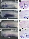
Expression of lamprey Gsc, Barx, Bapx, and Gdf5/6/7b in the embryonic and larval head. All larvae are Tahara st. 26.5 and are shown in side view (A, C, E, and G) and sagittal section (B, D, F, and H). (A and B) Gsc expression in the mesenchyme of the upper lip, first-arch gill bar (arrow), and distal lower lip (arrowhead). (C and D) Barx expression in the mesenchyme of the lower lip, extending dorsally into the ventral portion of the first gill bar (arrowhead). (E and F) Bapx expression in the epidermis of the ventral pharynx (arrowhead). (G and H) Gdf5/6/7b expression in the upper and lower lip ectoderm, upper lip mesenchyme, pharyngeal endoderm, ventral ectoderm ,and ventral mesenchyme surrounding the endostyle (arrowhead).
Fig. 3.
Fig. 3.
Comparison of DV patterning gene expression in the developing zebrafish and lamprey pharyngeal skeletons. (A) Ventral view of a 48hpf zebrafish embryo showing Bapx expression flanking the mouth in the forming jaw joints. (B) Lamprey Bapx expression is never observed in the nascent head skeleton. Dotted line marks the posterior edge of the mouth in both A and B. (C) DV extent of Gdf5, Bapx, Barx, Gsc, Dlx2,3,5,6, Msx, and Hand in the pharyngeal arches of the zebrafish. First-arch–specific patterning is depicted by the colored bars to the left of the diagram. Asterisk marks the jaw joint. (D) DV extent of Barx, Gsc, DlxA,B,C,D, Msx, Hand, and Gdf5/6/7b in the pharyngeal arches of the sea lamprey. First-arch–specific patterning is depicted by the colored bars to the left of the diagram. In both zebrafish and lamprey, combinatorial expression of Dlx, Msx, and Hand genes defines four domains of CNCC in the pharynx; ventral (v), ventral-intermediate (vi), dorsal-intermediate (di), and dorsal (d), suggesting sophisticated DV patterning is a basal feature of vertebrate head skeleton development. Although lamprey possesses a region topologically homologous to the jaw joint (asterisk), as indicated by similar first-arch–specific expression of Dlx, Msx, Hand, and Gsc, first-arch expression of Gdf5 and Bapx appears to be unique to gnathostomes, suggesting that recruitment of these genes to the first arch was associated with the evolution of the jaw. Combinatorial expression of lamprey Dlx, Msx, and Hand in the posterior pharynx corresponds to four distinct skeletal cell types, namely, mucocartilage (yellow), hypobranchial chondrocytes (green), “stack-of-coins” gill bar chondrocytes (light blue), and subchordal chondrocytes (dark blue). Pm, premandibular mesenchyme.

References

    1. Gans C, Northcutt RG. Neural crest and the origin of vertebrates: A new head. Science. 1983;220:268–273. - PubMed
    1. Clouthier DE, Schilling TF. Understanding endothelin-1 function during craniofacial development in the mouse and zebrafish. Birth Defects Res C Embryo Today. 2004;72:190–199. - PubMed
    1. Walshe J, Mason I. Fgf signalling is required for formation of cartilage in the head. Dev Biol. 2003;264:522–536. - PubMed
    1. Thomas T, et al. A signaling cascade involving endothelin-1, dHAND and msx1 regulates development of neural-crest-derived branchial arch mesenchyme. Development. 1998;125:3005–3014. - PubMed
    1. Miller CT, Yelon D, Stainier DY, Kimmel CB. Two endothelin 1 effectors, hand2 and bapx1, pattern ventral pharyngeal cartilage and the jaw joint. Development. 2003;130:1353–1365. - PubMed

Publication types

MeSH terms

Substances