

Plasma from preeclamptic women increases blood-brain barrier permeability: role of vascular endothelial growth factor signaling
- PMID: 20855653
- PMCID: PMC2959130
- DOI: 10.1161/HYPERTENSIONAHA.110.158931
Plasma from preeclamptic women increases blood-brain barrier permeability: role of vascular endothelial growth factor signaling
Abstract
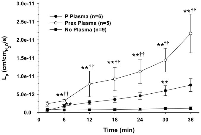
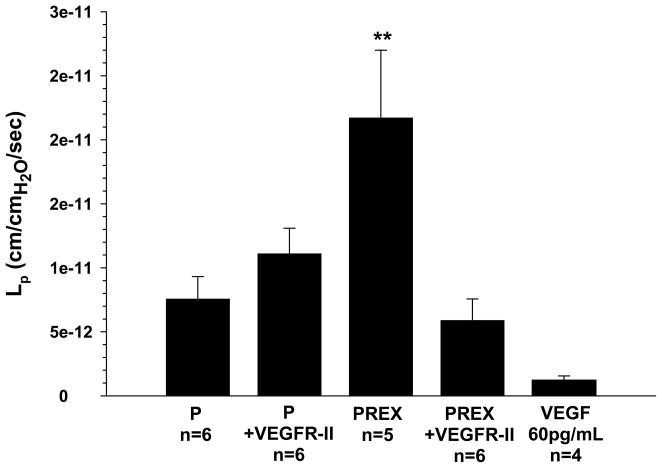
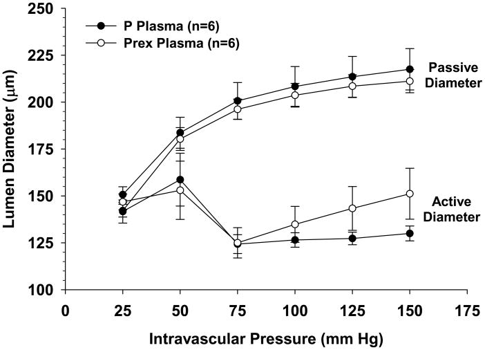
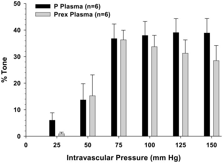
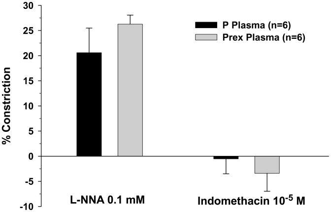
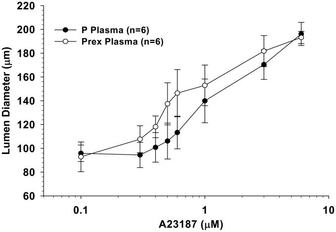
Circulating factors in preeclamptic women are thought to cause endothelial dysfunction and thereby contribute to the progression of this hypertensive condition. Despite the involvement of neurological complications in preeclampsia, there is a paucity of data regarding the effect of circulating factors on cerebrovascular function. Using a rat model of pregnancy, we investigated blood-brain barrier permeability, myogenic activity, and the influence of endothelial vasodilator mechanisms in cerebral vessels exposed intraluminally to plasma from normal pregnant or preeclamptic women. In addition, the role of vascular endothelial growth factor signaling in mediating changes in permeability in response to plasma was investigated. A 3-hour exposure to 20% normal pregnant or preeclamptic plasma increased blood-brain barrier permeability by ≈6.5- and 18.0-fold, respectively, compared with no plasma exposure (P<0.01). Inhibition of vascular endothelial growth factor receptor kinase activity prevented the increase in permeability in response to preeclamptic plasma but had no effect on changes in permeability of vessels exposed to normal pregnant plasma. Circulating factors in preeclamptic plasma did not affect myogenic activity or the influence of endothelium on vascular tone. These findings demonstrate that acute exposure to preeclamptic plasma has little effect on reactivity of cerebral arteries but significantly increases blood-brain barrier permeability. Prevention of increased permeability by inhibition of vascular endothelial growth factor signaling suggests that activation of this pathway may be responsible for increased blood-brain barrier permeability after exposure to preeclamptic plasma.
Conflict of interest statement
None.
Figures
References
-
- Duley L. The global impact of pre-eclampsia and eclampsia. Semin Perinatol. 2009;33:130–137. - PubMed
-
- Conrad KP, Benyo DF. Placental cytokines and the pathogenesis of preeclampsia. Am J Reprod Immunol. 1997;37:240–249. - PubMed
-
- Gilbert JS, Ryan MJ, LaMarca BB, Sedeek M, Murphy SR, Granger JP. Pathophysiology of hypertension during preeclampsia: linking placental ischemia with endothelial dysfunction. Am J Physiol Heart Circ Physiol. 2008;294:H541–H550. - PubMed
-
- Granger JP, Alexander BT, Llinas MT, Bennett WA, Khalil RA. Pathophysiology of preeclampsia: linking placental ischemia/hypoxia with microvascular dysfunction. Microcirculation. 2002;9:147–160. - PubMed
-
- Levine RJ, Maynard SE, Qian C, Lim KH, England LJ, Yu KF, Schisterman EF, Thadhani R, Sachs BP, Epstein FH, Sibai BM, Sukhatme VP, Karumanchi SA. Circulating angiogenic factors and the risk of preeclampsia. N Engl J Med. 2004;350:672–683. - PubMed
Publication types
MeSH terms
Substances
Grants and funding
LinkOut - more resources
Full Text Sources
Other Literature Sources
