. 2010 Oct 5;122(14):1364-72.
doi: 10.1161/CIRCULATIONAHA.110.945709. Epub 2010 Sep 20.

Noninvasive characterization of epicardial activation in humans with diverse atrial fibrillation patterns

Affiliations

Noninvasive characterization of epicardial activation in humans with diverse atrial fibrillation patterns

Phillip S Cuculich et al. Circulation. .

Abstract

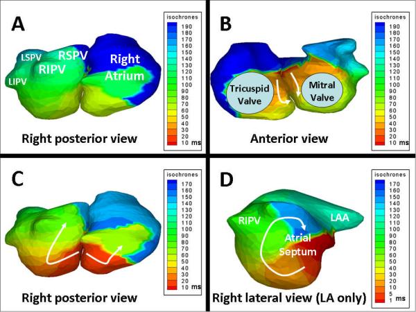
Background: Various mechanisms of atrial fibrillation (AF) have been demonstrated experimentally. Invasive methods to study these mechanisms in humans have limitations, precluding continuous mapping of both atria with sufficient resolution. In this article, we present continuous biatrial epicardial activation sequences of AF in humans using noninvasive electrocardiographic imaging (ECGI).

Methods and results: In the testing phase, ECGI accuracy was evaluated by comparing ECGI with co-registered CARTO images during atrial pacing in 6 patients. Additionally, correlative observations from catheter mapping and ablation were compared with ECGI in 3 patients. In the study phase, ECGI maps during AF in 26 patients were analyzed for mechanisms and complexity. ECGI noninvasively imaged the low-amplitude signals of AF in a wide range of patients (97 procedural success). Spatial accuracy for determining initiation sites from pacing was 6 mm. Locations critical to maintenance of AF identified during catheter ablation were identified by ECGI; ablation near these sites restored sinus rhythm. In the study phase, the most common patterns of AF were multiple wavelets (92), with pulmonary vein (69) and non-pulmonary vein (62) focal sites. Rotor activity was seen rarely (15). AF complexity increased with longer clinical history of AF, although the degree of complexity of nonparoxysmal AF varied widely.

Conclusions: ECGI offers a noninvasive way to map epicardial activation patterns of AF in a patient-specific manner. The results highlight the coexistence of a variety of mechanisms and variable complexity among patients. Overall, complexity generally increased with duration of AF.


Figures

Figure 1
Figure 1. ECGI accuracy locating atrial initiation sites, simulated by pacing
TOP LEFT: Isochrone map of LA (anterior view) with white star indicating earliest activation imaged by ECGI. BOTTOM LEFT: Potential map of LA locating the minimum of earliest activation (blue region, white star). TOP RIGHT: Merged 3-D CARTO and ECGI LA images (anterior view). Pacing sites in RSPV, atrial septum, and anterior mitral valve annulus are shown from CARTO (green circles) and ECGI (yellow diamonds). BOTTOM RIGHT: Detailed information regarding pacing sites and distance between CARTO and ECGI-imaged locations. RSPV=Right Superior Pulmonary Vein; RIPV=Right Inferior Pulmonary Vein; LAA=Left Atrial Appendage; MV=Mitral Valve.
Figure 2
Figure 2. Noninvasive ECGI of AF using a critical isthmus in the posterior LA during AF ablation (Movie 2 in Online Supplement)
Panel A shows a posterior view of the atria with a red star marking the location of the ablation that terminated AF. Panels B and C show ECGI isochrone maps during AF at two separate time points immediately prior to successful ablation. For both images, a wavefront enters the posterior LA (white arrows) through a protected isthmus. Online Movie 2 demonstrates these patterns with continuous AF imaging. LSPV=Left Superior Pulmonary Vein; LIPV=Left Inferior Pulmonary Vein.
Figure 3
Figure 3. Noninvasive ECGI of atypical atrial flutter during AF ablation (Movie 3 in Online Supplement)
Panels A and B show ECGI isochrone maps of both atria (right posterior and anterior views) during one cycle of atypical flutter. Earliest activation (red) is on the atrial septum in anterior view. Arrows designate the direction of wavefront propagation inferiorly and posterior. Panel C shows a different cycle, and highlights the posterior aspect of the septal reentry circuit. Panel D shows LA alone in right lateral view during flutter. The circus pattern of reentry is clearly seen along the atrial septum (arrow). Wavelets which do not participate in this circuit are present, likely causing variable activation patterns detected by invasive mapping.
Figure 4
Figure 4. Example of left PV focal sites in a patient with paroxysmal AF (Movie 4 in Online Supplement)
Panels A and B show two examples of ECGI isochrone maps of both atria (posterior view) at select times during AF. A focal site is frequently seen near the left PV's (stars) with radial activation spread. Conduction delay (crowded isochrones) is seen in posterior LA. Online Movie 4 demonstrates these patterns with continuous AF imaging, with 1 to 2 simultaneous wavelets predominantly traveling left-to-right.
Figure 5
Figure 5. Example of rotor pattern and focal site involving RIPV in a patient with persistent AF (Movie 5 in Online Supplement)
Panel A shows a reference image of both atria in posterior view. Panel B shows six time-lapse ECGI images of activation wavefronts (red) in a rotor pattern using RIPV as a pivot. White arrows show the path of wavefronts down the posterior LA and around the RIPV. At 50ms, activation exits the RIPV in a radial pattern. Online Movie 5 shows two to three simultaneous wavelets, frequently using RIPV as a pivot.
Figure 6
Figure 6. Example of single wave biatrial reentry in a patient with paroxysmal AF (Movie 6 in Online Supplement)
ECGI isochrone map of both atria in the right posterior and anterior views during 100ms of AF demonstrates a single spiral wave. The broad, sweeping activation wavefront involves both atria and propagates predominantly in a counterclockwise fashion (white arrows, anterior view). Although the surface ECG did not demonstrate clear regularity, this pattern on ECGI was highly repetitive. Black line marks the atrial septum. The Online Supplement Movie demonstrates this repetitive pattern.
Figure 7
Figure 7. Examples of complex rotor physiology in posterior LA in a patient with long-standing persistent AF (Movie 7 in Online Supplement)
Panel A: Activation pattern during AF (46–73ms) of both atria (posterior view). Activation wavelets are shown in red. White arrows indicate propagation direction of the wavelet. The white stars denote pivot points of wavelet rotation. At 46ms, a focal site emerges from LSPV and triggers a wave of radial activation (51ms). The emerging wavelet pivots around an area in the LA posterior wall (59–63ms) (star) and then propagates towards the right PVs (63–73ms). Panel B: Activation pattern during AF at a different time point. A wavelet at 503ms breaks into two on the posterior LA wall (508–525ms) and propagates around two pivot points (stars). At 533ms, the wavelets coalesce and terminate.
Figure 8
Figure 8. AF Complexity
Panel A: Increasing complexity of atrial fibrillation stratified by clinical classification of paroxysmal, persistent and longstanding persistent AF. With increasing AF duration, ECGI imaged more focal sites and wavelets (ANOVA for wavelet, p = 0.011; focal sites, p = 0.031). Panel B. Complexity Index is the sum of mean number of wavelets and number of focal sites for each patient. A Complexity Index of 1 is the minimum, and represents “simplest” AF. The Complexity Index increases from paroxysmal to persistent to longstanding persistent clinical groups. There is overlap in complexity between persistent and longstanding persistent groups.

References

    1. Stewart S, Hart CL, Hole DJ, McMurray JJ. A population-based study of the long-term risks associated with atrial fibrillation: 20-year follow-up of the Renfrew/Paisley study. Am J Med. 2002;113:359–64. - PubMed
    1. Wattigney WA, Mensah GA, Croft JB. Increasing trends in hospitalization for atrial fibrillation in the United States, 1985 through 1999. Circulation. 2003;108:711–6. - PubMed
    1. Fuster V, Ryden LE, Cannom DS, Crijns HJ, Curtis AB, Ellenbogen KA, Halperin JL, Le Heuzey JY, Kay GN, Lowe JE, Olsson SB, Prystowsky EN, Tamargo JL, Wann S, Smith SC, Jr., Jacobs AK, Adams CD, Anderson JL, Antman EM, Hunt SA, Nishimura R, Ornato JP, Page RL, Riegel B, Priori SG, Blanc JJ, Budaj A, Camm AJ, Dean V, Deckers JW, Despres C, Dickstein K, Lekakis J, McGregor K, Metra M, Morais J, Osterspey A, Zamorano JL. ACC/AHA/ESC 2006 Guidelines for the Management of Patients with Atrial Fibrillation. Circulation. 2006;114:e257–354. - PubMed
    1. Jalife J, Berenfeld O, Mansour M. Mother rotors and fibrillatory conduction: a mechanism of atrial fibrillation. Cardiovasc Res. 2002;54:204–16. - PubMed
    1. Chen YJ, Chen SA. Electrophysiology of pulmonary veins. J Cardiovasc Electrophysiol. 2006;17:220–4. - PubMed

Publication types