. 2010 Oct 15;185(8):4724-8.
doi: 10.4049/jimmunol.1001802. Epub 2010 Sep 20.

Acute ablation of Langerhans cells enhances skin immune responses

Affiliations

Acute ablation of Langerhans cells enhances skin immune responses

Aleh Bobr et al. J Immunol. .

Abstract

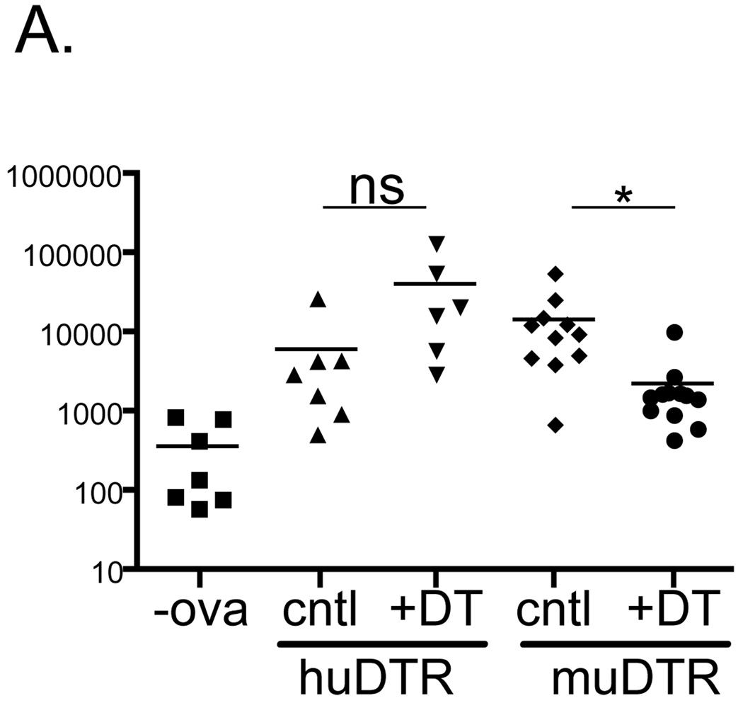
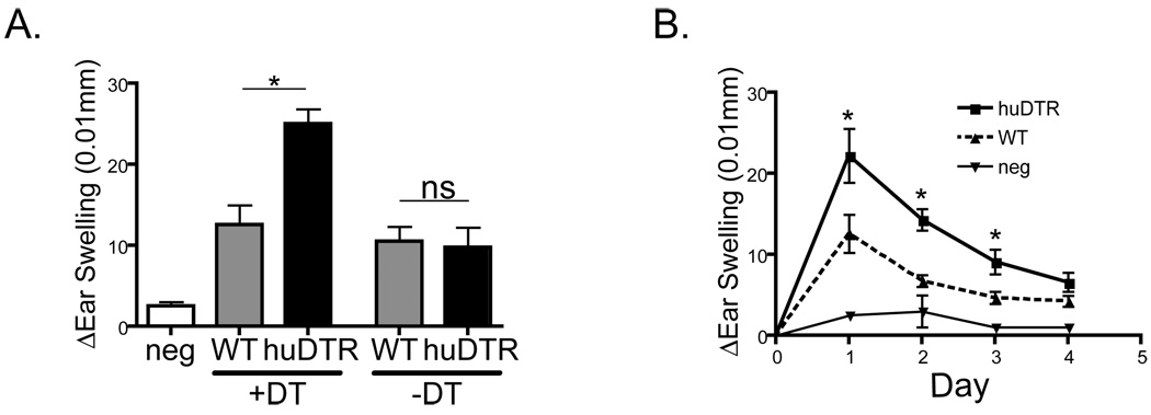
Understanding the function of Langerhans cells (LCs) in vivo has been complicated by conflicting results from LC-deficient mice. Human Langerin-DTA mice constitutively lack LCs and develop exaggerated contact hypersensitivity (CHS) responses. Murine Langerin-diphtheria toxin receptor (DTR) mice allow for the inducible elimination of LCs and Langerin(+) dermal dendritic cells (dDCs) after administration of diphtheria toxin, which results in reduced CHS. When Langerin(+) dDCs have partially repopulated the skin but LCs are still absent, CHS returns to normal. Thus, LCs appear to be suppressive in human Langerin-DTA mice and redundant in murine Langerin-DTR mice. To determine whether inducible versus constitutive LC ablation explains these results, we engineered human Langerin-DTR mice in which diphtheria toxin ablates LCs without affecting Langerin(+) dDCs. The inducible ablation of LCs in human Langerin-DTR mice resulted in increased CHS. Thus, LC-mediated suppression does not require their absence during ontogeny or during the steady-state and is consistent with a model in which LCs actively suppress Ag-specific CHS responses.


Figures

Figure 1
Figure 1. DTR expression in huDTR mice is specific for LC and allows for efficient ablation
A) Targeting construct for homologous recombination introducing the diphtheria toxin receptor (DTR) into the 3’UTR of langerin in human BAC RP11-504O1. B) Flow cytometry of epidermal single cell suspension from untreated huDTR mice stained with MHC II and CD45 to identify LC (CD45+, MHC-II+) and DETC (CD45+, MHC-II−). C) huLangerin expression in LC as gated in (B) in huDTR mice (solid line) and littermate control (shaded). D) As in (B), huDTR mice 2 days after DT administration. E) Percentage of LC and DETC among total epidermal cells in huDTR or control mice untreated or 2 days after DT administration, as specified. F) Immunofluorescence of epidermal whole mounts stained for MHC-II (red) or CD3 (green) from huDTR mice untreated or 2 days after DT administration. All data is representative of at least 3 independent experiments.
Figure 2
Figure 2. DT ablates LC leaving other Langerin+ DC unaffected
A) Flow cytometry of dermal single cell suspensions showing expression of CD11b and CD103 from WT, huDTR, and muDTR mice 2 days after DT administration. Cells were gated on muLangerin+, CD11c+. Numbers represent the percentage of cells in the indicated gate. B) Cutaneous LN (CLN), spleen and thymus from huDTR + DT, huDTR –DT and WT+DT stained for huLangerin and CD11c. C) CLN from untreated huDTR mice gated on muLangerin showing expression of huLangerin, CD103 and CD8. D) The depletion of huLangerin+ cells from epidermis and CLN at the indicated times after DT administration normalized to littermate controls. All data is representative of at least 3 independent experiments.
Figure 3
Figure 3. Langerin+ dDC are functionally intact in huDTR mice
2.5x10⁵ CD90.1 OT-I CD8 T cells were adoptive transferred into huDTR, muDTR and control mice. One day after administration of DT, mice were epicutaneouly immunized with 500 µg ovalbumin or vehicle (-ova). Five days later the number of OT-I cells from CLN was determined based on expression CD90.1. * p<0.05
Figure 4
Figure 4. Acute depletion of epidermal LC leads to enhanced CHS
A) WT and huDTR mice were sensitized with 0.5% DNFB on day+2 after DT or PBS administration. Five days later mice were challenged with 0.2% DNFB on the ear. Ear swelling one day after challenge (A) and over time (B) was measured. All data is representative of at least 3 independent experiments. * p<0.05

Similar articles

Cited by

References

    1. Banchereau J, Steinman RM. Dendritic cells and the control of immunity. Nature. 1998;392:245–252. - PubMed
    1. Romani N, Ebner S, Tripp CH, Flacher V, Koch F, Stoitzner P. Epidermal Langerhans cells--changing views on their function in vivo. Immunol Lett. 2006;106:119–125. - PubMed
    1. Kaplan DH, Kissenpfennig A, Clausen BE. Insights into Langerhans cell function from Langerhans cell ablation models. Eur J Immunol. 2008;38:2369–2376. - PubMed
    1. Kissenpfennig A, Henri S, Dubois B, Laplace-Builhe C, Perrin P, Romani N, Tripp CH, Douillard P, Leserman L, Kaiserlian D, Saeland S, Davoust J, Malissen B. Dynamics and Function of Langerhans Cells In Vivo Dermal Dendritic Cells Colonize Lymph Node AreasDistinct from Slower Migrating Langerhans Cells. Immunity. 2005;22:643–654. - PubMed
    1. Bennett CL, van Rijn E, Jung S, Inaba K, Steinman RM, Kapsenberg ML, Clausen BE. Inducible ablation of mouse Langerhans cells diminishes but fails to abrogate contact hypersensitivity. J Cell Biol. 2005;169:569–576. - PMC - PubMed

Publication types