. 2010 Nov 19;285(47):36842-8.
doi: 10.1074/jbc.M110.170324. Epub 2010 Sep 20.

CXCR3-B can mediate growth-inhibitory signals in human renal cancer cells by down-regulating the expression of heme oxygenase-1

Affiliations

CXCR3-B can mediate growth-inhibitory signals in human renal cancer cells by down-regulating the expression of heme oxygenase-1

Dipak Datta et al. J Biol Chem. .

Abstract

The chemokine receptor CXCR3 may play a critical role in the growth and metastasis of tumor cells, including renal tumors. It has been shown that CXCR3 has two splice variants with completely opposite functions; CXCR3-A promotes cell proliferation, whereas CXCR3-B inhibits cell growth. We recently demonstrated that the expression of growth-promoting CXCR3-A is up-regulated, and the growth-inhibitory CXCR3-B is markedly down-regulated in human renal cancer tissues; and the overexpression of CXCR3-B in renal cancer cells can significantly inhibit cell proliferation. However, the growth-inhibitory signal(s) through CXCR3-B are not well characterized. Here, we investigated the effector molecule(s) involved in CXCR3-B-mediated signaling events. We found that the overexpression of CXCR3-B in human renal cancer cells (Caki-1) promoted cellular apoptosis as observed by FACS analysis through Annexin-V staining. To examine whether the overexpression of CXCR3-B could alter the expression of any apoptosis-related genes in renal cancer cells, we performed a protein array. We found that CXCR3-B overexpression significantly down-regulated the expression of antiapoptotic heme oxygenase-1 (HO-1). By utilizing a HO-1 promoter-luciferase plasmid, we showed that CXCR3-B-mediated down-regulation of HO-1 was controlled at the transcriptional level as observed by luciferase assay. We also demonstrated that the inhibition of HO-1 expression using siRNA promoted apoptosis of renal cancer cells. Finally, we observed that human renal cancer tissues expressing low amounts of CXCR3-B significantly overexpress HO-1 at both mRNA and protein level. Together, we suggest that the overexpression of CXCR3-B may prevent the growth of renal tumors through the inhibition of antiapoptotic HO-1.


Figures

FIGURE 1.
FIGURE 1.
Overexpression of CXCR3-B in human renal cancer cells promotes apoptosis. Caki-1 cells were transfected with either the CXCR3-B overexpression plasmid (1.0 μg) or the empty expression vector. Following 48 h of transfection (A) the CXCR3-B mRNA expression was checked by real-time PCR (the lower Ct value reflects overexpression of the gene), and (B) the CXCR3 (A + B) protein expression was measured by FACS analysis. C, the confluence of the cells was evaluated microscopically. D, the apoptotic index of the cells was determined by Annexin-V and propidium iodide staining using an Apoptosis Detection kit as described under “Experimental Procedures.” All results are representative of three independent experiments.
FIGURE 2.
FIGURE 2.
Overexpression of CXCR3-B in renal cancer cells inhibits the expression of antiapoptotic HO-1. Caki-1 cells were transfected with either the CXCR3-B overexpression plasmid (1. 0 μg) or the empty expression vector for 48 h. The cells were lysed, and a protein array was performed utilizing an apoptosis protein array kit. The table chart beside the blot indicates the individual proteins tested in this array. Results are representative of three independent experiments.
FIGURE 3.
FIGURE 3.
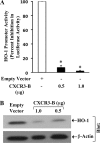
Overexpression of CXCR3-B inhibits HO-1 promoter activity. A, Caki-1 cells were co-transfected with the HO-1 promoter-luciferase construct (1.0 μg) and either different amounts (0.5 and 1.0 μg) of CXCR3-B overexpression plasmid (filled columns) or the empty expression vector (open column). The cells were harvested after 48 h, and the percent decrease in luciferase activity was calculated from luciferase counts of each group of cells compared with that of cells transfected with empty expression vector alone. Results are representative of three independent experiments. Columns, average of triplicate readings of samples; error bars, ± S.D. *, p < 0.01, compared with empty vector-transfected cells. B, Caki-1 cells were transfected with either different amounts (0.5 and 1.0 μg) of CXCR3-B overexpression plasmid or the empty expression vector. Following 48 h of transfection, whole cell lysates were prepared, and Western blot analysis was performed using anti-HO-1 to quantitate HO-1 protein expression. The expression of β-actin in these cells was analyzed by Western blotting using anti-β-actin. Results are representative of three independent experiments.
FIGURE 4.
FIGURE 4.
Down-regulation of HO-1 promotes apoptosis of renal cancer cells. Caki-1 cells were transfected with either the control or the HO-1 siRNA (50 nm) and cultured for 48 h. A, the apoptotic index of the cells was determined by Annexin-V and propidium iodide staining using an Apoptosis Detection kit as described under “Experimental Procedures.” B, the knockdown of HO-1 was confirmed by Western blot analysis. Results are representative of three independent experiments.
FIGURE 5.
FIGURE 5.
Overexpression of HO-1 inhibits apoptosis of renal cancer cells. Caki-1 cells were treated with either CoPP (50 μm) or the vehicle alone for 48 h. A, apoptotic index of the cells was determined by Annexin-V and propidium iodide staining using an Apoptosis Detection kit as described under “Experimental Procedures.” B, overexpression of HO-1 following CoPP treatment was confirmed by Western blot analysis. Results are representative of three independent experiments.
FIGURE 6.
FIGURE 6.
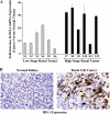
HO-1 is overexpressed in human renal cancer tissues. A, total RNA was isolated from renal cancer and normal renal tissues and reverse transcribed. The fold increase of HO-1 mRNA expression in renal cancer tissues versus normal renal tissues was measured by real-time PCR. S1–S6 represent low stage (Robson stages I and II), whereas S7–S12 represent high stage (Robson stages III and IV) renal tumor tissues. Columns are average of triplicate readings of the samples; error bars are ± S.D. B, representative photomicrographs show the expression of HO-1 in human renal cell cancer and normal kidney tissues detected by immunohistochemistry. Magnification, ×400. Brown color, expression of HO-1. Results are representative of three different tissues.

References

    1. Zlotnik A., Yoshie O. (2000) Immunity 12, 121–127 - PubMed
    1. Balkwill F., Mantovani A. (2001) Lancet. 357, 539–545 - PubMed
    1. Proudfoot A. E. (2002) Nat. Rev. Immunol. 2, 106–115 - PMC - PubMed
    1. Zlotnik A. (2004) Semin Cancer Biol. 14, 181–185 - PubMed
    1. Strieter R. M., Belperio J. A., Phillips R. J., Keane M. P. (2004) Semin Cancer Biol. 14, 195–200 - PubMed

Publication types

MeSH terms