

Epimorphic regeneration in mice is p53-independent
- PMID: 20855943
- PMCID: PMC3047795
- DOI: 10.4161/cc.9.18.13119
Epimorphic regeneration in mice is p53-independent
Abstract
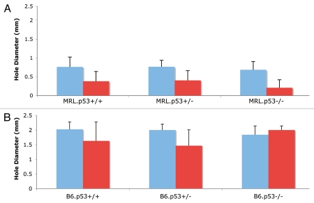
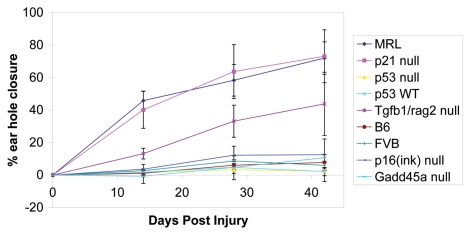
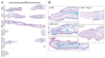
The process of regeneration is most readily studied in species of sponge, hydra, planarian and salamander (i.e., newt and axolotl). The closure of MRL mouse ear pinna through-and-through holes provides a mammalian model of unusual wound healing/regeneration in which a blastema-like structure closes the ear hole and cartilage and hair follicles are replaced. Recent studies, based on a broad level of DNA damage and a cell cycle pattern of G₂/M "arrest," showed that p21(Cip1/Waf1) was missing from the MRL mouse ear and that a p21-null mouse could close its ear holes. Given the p53/p21 axis of control of DNA damage, cell cycle arrest, apoptosis and senescence, we tested the role of p53 in the ear hole regenerative response. Using backcross mice, we found that loss of p53 in MRL mice did not show reduced healing. Furthermore, cross sections of MRL. p53(-/-) mouse ears at 6 weeks post-injury showed an increased level of adipocytes and chondrocytes in the region of healing whereas MRL or p21(-/-) mice showed chondrogenesis alone in this same region, though at later time points. In addition, we also investigated other cell cycle-related mutant mice to determine how p21 was being regulated. We demonstrate that p16 and Gadd45 null mice show little healing capacity. Interestingly, a partial healing phenotype in mice with a dual Tgfβ/Rag2 knockout mutation was seen. These data demonstrate an independence of p53 signaling for mouse appendage regeneration and suggest that the role of p21 in this process is possibly through the abrogation of the Tgfβ/Smad pathway.
Figures
Similar articles
-
Lack of p21 expression links cell cycle control and appendage regeneration in mice.Proc Natl Acad Sci U S A. 2010 Mar 30;107(13):5845-50. doi: 10.1073/pnas.1000830107. Epub 2010 Mar 15. Proc Natl Acad Sci U S A. 2010. PMID: 20231440 Free PMC article.
-
The role of p21 in regulating mammalian regeneration.Stem Cell Res Ther. 2011 Jun 29;2(3):30. doi: 10.1186/scrt71. Stem Cell Res Ther. 2011. PMID: 21722344 Free PMC article.
-
Regeneration of the ear after wounding in different mouse strains is dependent on the severity of wound trauma.Dev Dyn. 2003 Feb;226(2):388-97. doi: 10.1002/dvdy.10242. Dev Dyn. 2003. PMID: 12557217
-
Induction of p21(Waf1/Cip1) by garcinol via downregulation of p38-MAPK signaling in p53-independent H1299 lung cancer.J Agric Food Chem. 2014 Mar 5;62(9):2085-95. doi: 10.1021/jf4037722. Epub 2014 Feb 24. J Agric Food Chem. 2014. PMID: 24533688
-
Spallanzani's mouse: a model of restoration and regeneration.Curr Top Microbiol Immunol. 2004;280:165-89. doi: 10.1007/978-3-642-18846-6_5. Curr Top Microbiol Immunol. 2004. PMID: 14594211 Review.
Cited by
-
Increased AMP-activated protein kinase in skeletal muscles of Murphy Roth Large mice and its potential role in altered metabolism.Physiol Rep. 2014 Mar 20;2(3):e00252. doi: 10.1002/phy2.252. Print 2014. Physiol Rep. 2014. PMID: 24760507 Free PMC article.
-
Tumor suppressors: enhancers or suppressors of regeneration?Development. 2013 Jun;140(12):2502-12. doi: 10.1242/dev.084210. Development. 2013. PMID: 23715544 Free PMC article.
-
Enhanced cartilage repair in 'healer' mice-New leads in the search for better clinical options for cartilage repair.Semin Cell Dev Biol. 2017 Feb;62:78-85. doi: 10.1016/j.semcdb.2016.04.018. Epub 2016 Apr 26. Semin Cell Dev Biol. 2017. PMID: 27130635 Free PMC article. Review.
-
The Role of TGFβ Signaling in Wound Epithelialization.Adv Wound Care (New Rochelle). 2014 Jul 1;3(7):482-491. doi: 10.1089/wound.2013.0466. Adv Wound Care (New Rochelle). 2014. PMID: 25032068 Free PMC article. Review.
-
Oxygen, Metabolism, and Regeneration: Lessons from Mice.Trends Mol Med. 2017 Nov;23(11):1024-1036. doi: 10.1016/j.molmed.2017.08.008. Epub 2017 Oct 5. Trends Mol Med. 2017. PMID: 28988849 Free PMC article. Review.
References
-
- Stocum DL. The urodele limb regeneration blastema. Determination and organization of the morphogenetic field. Differentiation. 1984;27:55–57. - PubMed
-
- Carlson BM. Some principles of regeneration in mammalian systems. 2010:4–13. - PubMed
-
- Kragl M, Knapp D, Nacu E, Khattak S, Maden M, Epperlein HH, et al. Cells keep a memory of their tissue origin during axolotl limb regeneration. Nature. 2009;460:60–65. - PubMed
-
- Kierdorf U, Kierdorf H. Deer antlers—a model of mammalian appendage regeneration: An extensive review. Gerontology. 2010 [Epub ahead of print] - PubMed
Publication types
MeSH terms
Substances
Grants and funding
LinkOut - more resources
Full Text Sources
Research Materials
Miscellaneous