. 2011 Jan 27;30(4):482-93.
doi: 10.1038/onc.2010.435. Epub 2010 Sep 20.

Human AP endonuclease (APE1/Ref-1) and its acetylation regulate YB-1-p300 recruitment and RNA polymerase II loading in the drug-induced activation of multidrug resistance gene MDR1

Affiliations

Human AP endonuclease (APE1/Ref-1) and its acetylation regulate YB-1-p300 recruitment and RNA polymerase II loading in the drug-induced activation of multidrug resistance gene MDR1

S Sengupta et al. Oncogene. .

Abstract

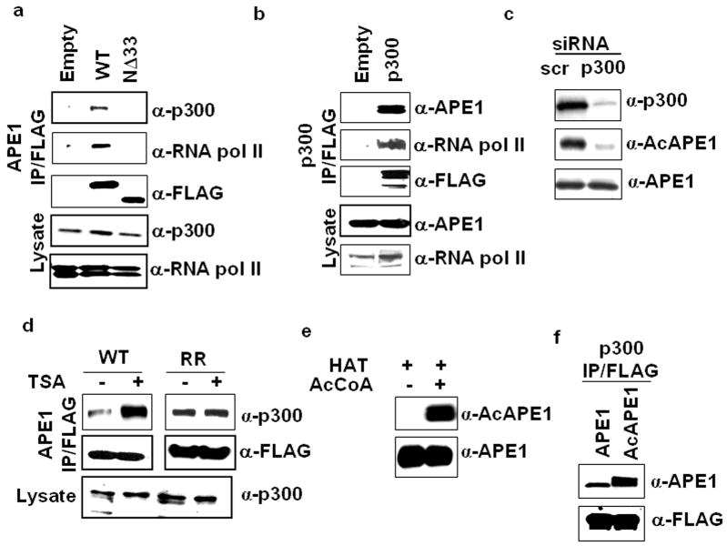
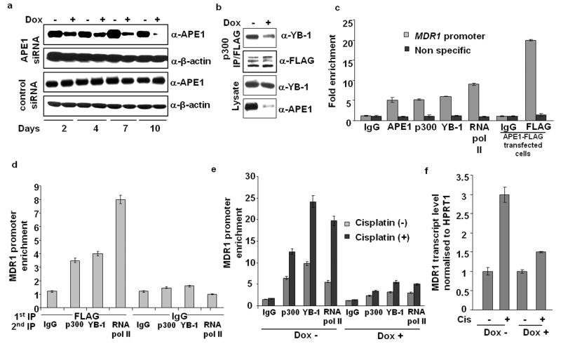
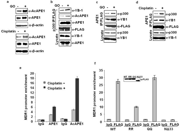
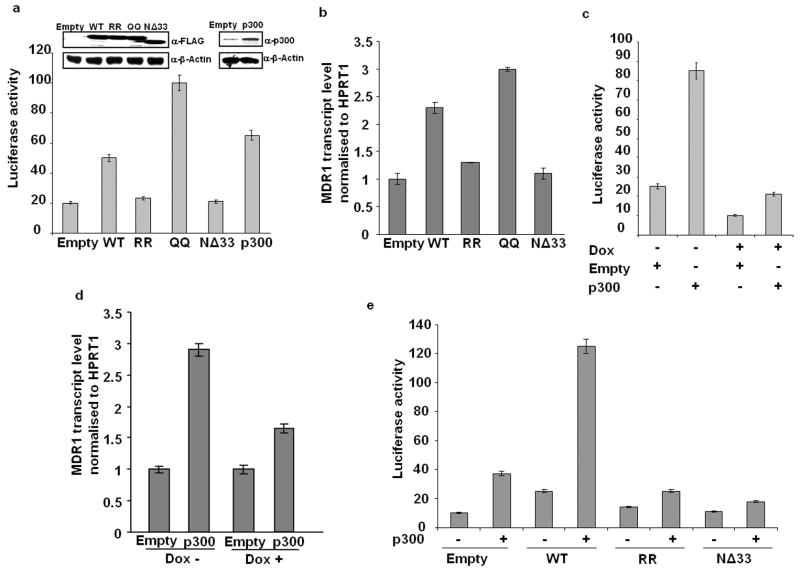
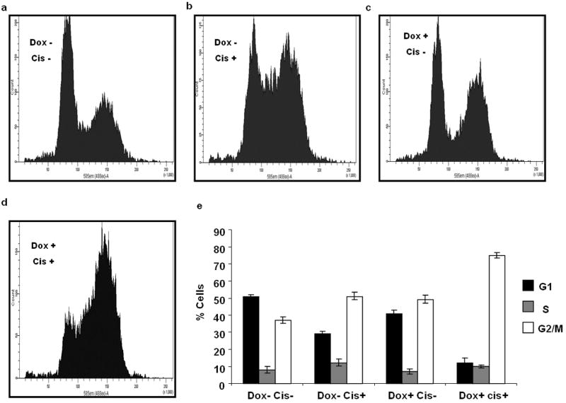
The overexpression of human apurinic/apyrimidinic (AP) endonuclease 1 (APE1/Ref-1), a key enzyme in the DNA base excision repair (BER) pathway, is often associated with tumor cell resistance to various anticancer drugs. In this study, we examined the molecular basis of transcriptional regulatory (nonrepair) function of APE1 in promoting resistance to certain types of drugs. We have recently shown that APE1 stably interacts with Y-box-binding protein 1 (YB-1), and acts as its coactivator for the expression of multidrug resistance gene MDR1, thereby causing drug resistance. In this study, we show, to the best of our knowledge, for the first time that APE1 is stably associated with the basic transcription factor RNA polymerase II (RNA pol II) and the coactivator p300 on the endogenous MDR1 promoter. The depletion of APE1 significantly reduces YB-1-p300 recruitment to the promoter, resulting in reduced RNA pol II loading. Drug-induced APE1 acetylation, which is mediated by p300, enhances formation of acetylated APE1 (AcAPE1)-YB-1-p300 complex on the MDR1 promoter. Enhanced recruitment of this complex increases MDR1 promoter-dependent luciferase activity and its endogenous expression. Using APE1-downregulated cells and cells overexpressing wild-type APE1 or its nonacetylable mutant, we have demonstrated that the loss of APE1's acetylation impaired MDR1 activation and sensitizes the cells to cisplatin or etoposide. We have thus established the basis for APE1's acetylation-dependent regulatory function in inducing MDR1-mediated drug resistance.


Conflict of interest statement

Conflict of Interest

The authors declare no potential conflict of interest.

Figures

Figure 1
Figure 1
Association of APE1 with p300 and RNA pol II. (a) Nuclear lysates of HEK-293T cells transfected with empty vector, FLAG-tagged WT APE1 or N terminal 33 aa deleted (NΔ33) APE1 mutant were immunoprecipitated (IP) with FLAG antibody. Western analysis of the IPs with p300, RNA pol II or FLAG antibody; p300 and RNA pol II levels in the lysates (lower panels). (b) Nuclear extracts of HEK-293T cells transfected with empty vector or FLAG tagged p300 were immunoprecipitated with FLAG antibody. Western analysis of the IPs with APE1, RNA pol II or FLAG antibody; APE1 and RNA pol II levels in the lysates (lower panels). (c) HEK-293T cells were transfected with 80 nM of p300–specific or scrambled (scr) duplex siRNAs. Western analysis after 48 hours for p300 (upper panel), AcAPE1 (middle panel) and APE1 (lower panel) levels in cell extracts. (d) HEK-293T cells transfected with FLAG-tagged WT APE1 (WT) or K6R/K7R APE1 (RR) were treated with TSA (100 ng/ml) for 6 hours and then the nuclear extracts were immunoprecipitated with FLAG antibody. Western analysis of the IPs with p300 (upper panel) or FLAG (middle panel) antibody; p300 levels in the lysates (lower panel). (e) Recombinant APE1 (2 μg) was incubated with p300 HAT domain in presence (+) or absence (-) of 1 mM acetyl CoA (AcCoA) for 2 hours at 30°C. Western analysis of the invitro acetylated APE1 with AcAPE1 (upper panel) or APE1 (lower panel) antibody. (f) Immunoprecipitated p300-FLAG beads were incubated with 100 ng of unmodified or invitro acetylated APE1. After washing, the bound proteins were eluted with SDS/Laemmli buffer and immunoblotted with APE1 (upper panel) or FLAG (lower panel) antibody.
Figure 2
Figure 2
Requirement of APE1 in YB-1/p300 recruitment and RNA pol II loading on MDR1 promoter. (a) HEK-293T cells expressing APE1 siRNA (APE1siRNAHEK-293T cells) or control duplex siRNA (ControlsiRNAHEK-293T cells) under a Doxycycline (Dox)-inducible promoter were established following the procedure described in Materials & Methods. These cells were treated with Dox (1ug/ml) for the indicated times and APE1 levels were measured by Western analysis; β-actin antibody was used as a loading control. (b) APE1siRNAHEK-293T cells were treated with (+) or without (-) Dox for 8 days and then the cells were transfected with p300-FLAG expression plasmid. Nuclear extracts were immunoprecipitated with FLAG antibody and Western analysis of the IPs was performed with YB-1 or FLAG antibody; YB-1 and APE1 levels in the lysates (lower panels). (c) CHIP assay in HEK-293T cells or APE1-FLAG transfected HEK-293T cells with the indicated antibodies. Immunoprecipitated Y-box element containing MDR1 promoter sequence or a non specific coding sequence in the MDR1 gene distant to Y-box element was amplified and quantified by SYBR GREEN based Real Time PCR analysis. Values in the bar diagram are relative to PCR from input chromatin. (d) re-ChIP assay of immunoprecipitated APE1-FLAG bound chromatin from APE1 FLAG transfected HEK-293T cells with p300, YB-1 or RNA pol II Ab or control IgG. The first IP was carried out with FLAG antibody or control IgG and the second IP was carried out with p300, YB-1 or RNA pol II Antibody or control IgG. (e) APE1 levels in APE1siRNAHEK-293T cells were downregulated with Dox treatment. Then the cells were treated with (+) or without (-) cisplatin for 1 hour, and 5 hours later, ChIP assay was carried out with p300, YB-1 or RNA pol II Antibody or control IgG. (f) Real Time RT-PCR analysis of MDR1 transcript. APE1 levels in APE1siRNAHEK-293T cells were downregulated with Dox treatment. The cells were then treated with cisplatin for 1 hour and 16 hours later total RNA was isolated and subjected to cDNA synthesis and Real Time PCR. Values in the bar diagram (normalized with respect to HPRT1 transcript) are relative to cisplatin untreated control. All the results represent the mean ± standard deviations of 3 independent experiments performed in duplicates.
Figure 3
Figure 3
Oxidative stress enhances APE1’s acetylation and its association with YB-1/p300 on MDR1 promoter. (a) HEK 293T cells were treated with 100 ng/ml of Glucose oxidase (GO) or 40 μg/ml of cisplatin for 1 hour, and 5 hours later the AcAPE1, APE1 or β-actin levels in the whole cell extracts were measured by Western analysis. (b) HEK-293T cells transfected with FLAG-tagged p300 were treated with GO (100 ng/ml) for 1 hour, and 5 hours later, nuclear lysates were prepared and immunoprecipitated with FLAG antibody. Western analysis of the IPs with YB-1, AcAPE1, APE1 or FLAG antibody; YB-1 and APE1 levels in the lysates (lower panels). (c & d) HEK-293T cells transfected with FLAG-tagged APE1 were treated with GO (100 ng/ml) or cisplatin (40 μg/ml) as above, nuclear lysates were prepared and immunoprecipitated with FLAG antibody. Western analysis of the IPs with p300, YB-1 or FLAG antibody; p300 and YB-1 levels in the lysates (lower panels). (e) ChIP assay for the association of AcAPE1 on MDR1 promoter. HEK-293T cells were treated with cisplatin as above and ChIP assay was performed with APE1 or AcAPE1 antibody or control IgG. (f) HEK-293T cells were transfected with WT APE1, RR, QQ or NΔ33 APE1 and ChIP assay was carried out with FLAG antibody or control IgG. Inset panel shows the level of FLAG tagged WT or mutant APE1 proteins in the cell extracts determined by Western analysis with FLAG antibody. Values in the bar diagram in all the cases are relative to PCR from input chromatin and results represent mean ± standard deviations of 3 independent ChIP experiments performed in duplicates.
Figure 4
Figure 4
Enhancement of MDR1 promoter activity and expression by APE1 acetylation. (a) HEK-293T cells were cotransfected with MDR1 promoter-luciferase reporter plasmid and empty vector, WT, RR, QQ, NΔ33 APE1 or p300 expression plasmids. 48 hours later, luciferase activity was measured and normalized with total protein content in the lysates. Inset panel shows the level of FLAG tagged WT/ mutant APE1 proteins or p300 protein in the cell extracts determined by Western analysis. (b) Real Time RT-PCR assay for MDR1 transcript levels in HEK-293T cells ectopically expressing WT, RR, QQ or NΔ33 APE1 mutant proteins. Values in the bar diagram are relative to empty vector transfected cells. (c) APE1 levels in APE1siRNAHEK-293T cells were downregulated with Dox treatment for 8 days. Then the cells were cotransfected with MDR1 promoter reporter plasmid and expression plasmids for p300 or its empty vector. Luciferase activity was measured as mentioned above. (d) MDR1 transcript levels in Dox-treated (APE1 downregulated) or untreated APE1siRNAHEK-293T cells ectopically expressing p300. Values in the bar diagram are relative to empty vector transfected cells. (e) HEK-293T cells were cotransfected with MDR1 promoter reporter plasmid along with WT, RR or NΔ33 APE1 constructs and p300 expression plasmid. Luciferase activity was measured as mentioned above. Results in all the cases represent the mean ± standard deviations of 3 independent experiments performed in duplicates.
Figure 5
Figure 5
APE1 depletion induces cisplatin-mediated G2/M arrest. (a-d) Cell cycle distribution pattern of APE1 downregulated and control APE1siRNAHEK-293T cells treated with cisplatin. After knockdown of endogenous APE1 level with Dox treatment for 8 days, the cells were treated with 100 μM cisplatin and after 24 hours the cells were analysed for FACS as described in Materials and Methods. (e) The percentage of cells at G1, S and G2/M stages were determined by FACSDiva Version 6.1.2 software programme and plotted and the results in each case represent the mean ± standard deviations of 3 independent experiments performed in duplicates.
Figure 6
Figure 6
Involvement of APE1’s acetylation-mediated regulatory function in resistance to cisplatin or etoposide. (a & c) Clonogenic survival assay of APE1siRNAHEK-293T cells treated with Dox. APE1 level in these cells was downregulated with Dox treatment for 8 days and seeded in 35 mm dishes (~ 300 cells/dish). Eighteen hours later, the cells were treated with increasing concentrations of cisplatin (0 to 125 μM) or etoposide (0 to 20 μM). Cells were maintained for 10 -14 days and the colonies were fixed and stained with giemsa stain. (b & d) After Dox treatment for 8 days, APE1siRNAHEK-293T cells were transfected with empty vector, WT APE1, RR APE1 or QQ APE1 expression plasmids. Forty eight hours later, the cells were seeded in 35 mm dishes (~ 300 cells/dish) and proceeded as above. The number of visible colonies was counted and one hundred percent corresponds to the number of colonies in the absence of drugs. The graph shows the mean ± standard deviations of 3 independent experiments performed in duplicates

References

    1. Ahmad S, Glazer RI. Expression of the antisense cDNA for protein kinase C alpha attenuates resistance in doxorubicin-resistant MCF-7 breast carcinoma cells. Mol Pharmacol. 1993;43:858–862. - PubMed
    1. Ahmad S, Safa AR, Glazer RI. Modulation of P-glycoprotein by protein kinase C alpha in a baculovirus expression system. Biochemistry. 1994;33:10313–10318. - PubMed
    1. Ambudkar SV, Sauna ZE, Gottesman MM, Szakacs G. A novel way to spread drug resistance in tumor cells: functional intercellular transfer of P-glycoprotein (ABCB1) Trends Pharmacol Sci. 2005;26:385–387. - PMC - PubMed
    1. Ando K, Hirao S, Kabe Y, Ogura Y, Sato I, Yamaguchi Y, et al. A new APE1/Ref-1-dependent pathway leading to reduction of NF-kappaB and AP-1, and activation of their DNA-binding activity. Nucleic Acids Res. 2008;36:4327–4336. - PMC - PubMed
    1. Bapat A, Fishel M, Kelley MR. Going Ape as an Approach to Cancer Therapeutics. Antioxid Redox Signal. 2009;11:651–668. - PMC - PubMed

Publication types

MeSH terms

Substances