

The role of clinical parapapillary atrophy evaluation in the diagnosis of open angle glaucoma
- PMID: 20856591
- PMCID: PMC2938276
- DOI: 10.2147/opth.s12420
The role of clinical parapapillary atrophy evaluation in the diagnosis of open angle glaucoma
Abstract
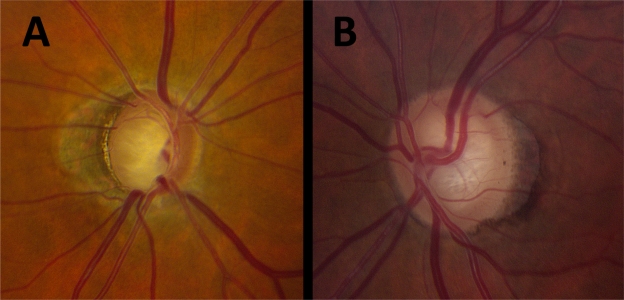
Purpose: To determine if clinical evaluation of parapapillary atrophy (PPA) significantly improves the ability to distinguish open-angle glaucoma (OAG) patients from glaucoma suspects.
Methods: Patients in this study were under evaluation for glaucoma and had open angles, at least one reliable 24-2 SITA-standard automatic perimetry, and digital stereophotographs of the optic disc. PPA was identified clinically as a parapapillary region of absent (βPPA) or hyper/hypopigmented (αPPA) retinal pigment epithelium. A single masked observer evaluated photos for: vertical cup-to-disc ratio (CDR), clock hours of total and βPPA, βPPA as percentage width of the optic disc, presence or absence of βPPA at each disc quadrant, and ordinal rating of total PPA. Generalized linear models were used to determine odds of an abnormal or borderline glaucoma hemifield test (GHT) as a function of PPA variables and covariates; model fit was assessed using the log-likelihood ratio test.
Results: Of 410 consecutive patients, 540 eyes (of 294 patients) met inclusion criteria. Mean age was greater among patients with abnormal compared with normal GHT (P < 0.001), but sex and race/ethnicity did not differ between groups (P ≥ 0.22). Age, central corneal thickness (CCT) and CDR (P ≤ 0.006), but not intraocular pressure (IOP) (P = 0.71), were significant univariable predictors of the odds of an abnormal GHT. All PPA parameters significantly predicted GHT (P ≤ 0.03), except presence of temporal βPPA (P = 0.25). Adjustment for age, CCT, IOP, and CDR reduced the association between PPA and GHT, and model fit was not greatly improved by addition of PPA variables.
Conclusions: Addition of most PPA parameters to a model already containing commonly assessed variables including age, CCT, IOP, and CDR does not significantly improve the ability to distinguish OAG patients from glaucoma suspects.
Keywords: glaucoma; optic nerve; parapapillary atrophy; visual fields.
Figures
Similar articles
-
Beta-Zone parapapillary atrophy and the velocity of glaucoma progression.Ophthalmology. 2010 May;117(5):909-15. doi: 10.1016/j.ophtha.2009.10.016. Epub 2010 Feb 4. Ophthalmology. 2010. PMID: 20132988
-
Association between Corneal Deformation Amplitude and Posterior Pole Profiles in Primary Open-Angle Glaucoma.Ophthalmology. 2016 May;123(5):959-64. doi: 10.1016/j.ophtha.2015.12.043. Epub 2016 Feb 11. Ophthalmology. 2016. PMID: 26875001
-
β-Zone Parapapillary Atrophy and Rates of Glaucomatous Visual Field Progression: African Descent and Glaucoma Evaluation Study.JAMA Ophthalmol. 2017 Jun 1;135(6):617-623. doi: 10.1001/jamaophthalmol.2017.1082. JAMA Ophthalmol. 2017. PMID: 28494060 Free PMC article.
-
Anatomic relationships between disc hemorrhage and parapapillary atrophy.Am J Ophthalmol. 2008 Nov;146(5):735-40. doi: 10.1016/j.ajo.2008.06.018. Epub 2008 Aug 23. Am J Ophthalmol. 2008. PMID: 18723142
-
Optic Disc Change during Childhood Myopic Shift: Comparison between Eyes with an Enlarged Cup-To-Disc Ratio and Childhood Glaucoma Compared to Normal Myopic Eyes.PLoS One. 2015 Jul 6;10(7):e0131781. doi: 10.1371/journal.pone.0131781. eCollection 2015. PLoS One. 2015. PMID: 26147983 Free PMC article.
Cited by
-
Controversies in the association of parapapillary atrophy with glaucoma.Taiwan J Ophthalmol. 2019 Oct 31;10(4):243-249. doi: 10.4103/tjo.tjo_64_19. eCollection 2020 Oct-Dec. Taiwan J Ophthalmol. 2019. PMID: 33437595 Free PMC article. Review.
-
Comparative analysis of OCT-defined parapapillary beta and gamma zones between primary open angle glaucoma and primary angle closure glaucoma.Sci Rep. 2022 Jun 30;12(1):11070. doi: 10.1038/s41598-022-15457-3. Sci Rep. 2022. PMID: 35773326 Free PMC article.
-
Longitudinal changes in peripapillary atrophy in the ocular hypertension treatment study: a case-control assessment.Ophthalmology. 2015 Jan;122(1):79-86. doi: 10.1016/j.ophtha.2014.07.033. Epub 2014 Sep 7. Ophthalmology. 2015. PMID: 25208858 Free PMC article.
-
A novel glaucomatous representation method based on Radon and wavelet transform.BMC Bioinformatics. 2019 Dec 24;20(Suppl 25):693. doi: 10.1186/s12859-019-3267-6. BMC Bioinformatics. 2019. PMID: 31874641 Free PMC article.
-
Peripapillary atrophy classification using CNN deep learning for glaucoma screening.PLoS One. 2022 Oct 6;17(10):e0275446. doi: 10.1371/journal.pone.0275446. eCollection 2022. PLoS One. 2022. PMID: 36201448 Free PMC article.
References
-
- Harper R, Reeves B. The sensitivity and specificity of direct ophthalmoscopic optic disc assessment in screening for glaucoma: a multivariate analysis. Graefes Arch Clin Exp Ophthalmol. 2000;238(12):949–955. - PubMed
-
- Theodossiades J, Murdoch I. What optic disc parameters are most accurately assessed using the direct ophthalmoscope? Eye (Lond) 2001;15(Pt 3):283–287. - PubMed
-
- Jonas JB, Budde WM, Panda-Jonas S. Ophthalmoscopic evaluation of the parapapillary region of the optic nerve head. Klin Oczna. 2004;106(Suppl 1–2):279–289. - PubMed
-
- Jonas JB, Naumann GO. Parapapillary chorioretinal atrophy in normal and glaucoma eyes. II. Correlations. Invest Ophthalmol Vis Sci. 1989;30(5):919–926. - PubMed
LinkOut - more resources
Full Text Sources
Miscellaneous
