

Intentional sparing of daughter sac from coil packing in the embolization of aneurysms causing the third cranial nerve palsy : initial clinical and radiological results
- PMID: 20856658
- PMCID: PMC2941852
- DOI: 10.3340/jkns.2010.48.2.115
Intentional sparing of daughter sac from coil packing in the embolization of aneurysms causing the third cranial nerve palsy : initial clinical and radiological results
Abstract
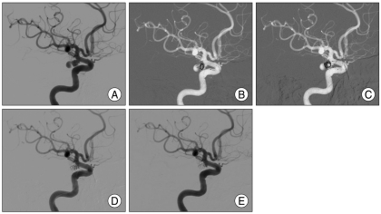
Objective: Cerebral aneurysms which cause oculomotor nerve [cranial nerve (CN) III] palsy, are frequently found with a daughter sac of the aneurysm dome. We assumed that CN III might be compressed by the daughter sac and it would be more helpful not to fill the daughter sac with coils than vice versa during endosaccular embolization for recovering from CN III palsy, because it may give a greater chance for the daughter sac to shrink by itself later. We reviewed the initial results of our experiences of such cases.
Methods: Among 9 aneurysms accompanied by CN III palsy, 7 (6 unruptured, 1 ruptured) showed a daughter sac. We tried to fill the main dome completely and spare the daughter sac from coil filling to increase the possibility of decompression. We evaluated the short-term effectiveness of this concept using medical records and angiograms.
Results: After initial embolization, all of CN III palsy caused by unruptured aneurysms (6/6) resolved completely after various periods (3-90 days) of time. No adverse effects were noted during and after the procedures except for one case of harmless coil stretching during coil filling using double microcatheter.
Conclusion: During the coil embolization of the cerebral aneurysm causing CN III palsy, sparing the daughter sac from coil packing while tightly packing the main dome, can be helpful in increasing the effectiveness of decompression. However, a long-term follow-up will be required.
Keywords: Cerebral aneurysm; Decompression; Embolization; Oculomotor nerve palsy.
Figures

Similar articles
-
Isolated abducens nerve palsy associated with subarachnoid hemorrhage: a localizing sign of ruptured posterior inferior cerebellar artery aneurysms.J Neurosurg. 2018 Jun;128(6):1830-1838. doi: 10.3171/2017.2.JNS162951. Epub 2017 Sep 1. J Neurosurg. 2018. PMID: 28862551
-
Unruptured aneurysms with cranial nerve symptoms: efficacy of endosaccular Guglielmi detachable coil treatment.Korean J Radiol. 2003 Jul-Sep;4(3):141-5. doi: 10.3348/kjr.2003.4.3.141. Korean J Radiol. 2003. PMID: 14530641 Free PMC article.
-
Early ceasing of intra-aneurysmal contrast opacification during coil embolization in ruptured aneurysms compared with unruptured aneurysms.Neurosurgery. 2011 Sep;69(3):651-8; discussion 658. doi: 10.1227/NEU.0b013e31821bc4b4. Neurosurgery. 2011. PMID: 21499153
-
[Coil Embolization for Very Small Intracranial Aneurysms with Diameter Less than 3mm:A Case Series of 14 Patients and Literature Review].No Shinkei Geka. 2018 Apr;46(4):303-312. doi: 10.11477/mf.1436203721. No Shinkei Geka. 2018. PMID: 29686163 Review. Japanese.
-
Cranial nerve palsies and intracranial aneurysms: A narrative review of patterns and outcomes.Surg Neurol Int. 2024 Aug 9;15:277. doi: 10.25259/SNI_531_2024. eCollection 2024. Surg Neurol Int. 2024. PMID: 39246770 Free PMC article. Review.
Cited by
-
Endovascular Treatment of Patients with Oculomotor Nerve Palsy Induced by Posterior Communicating Artery Aneurysms.J Neuroendovasc Ther. 2020;14(9):366-372. doi: 10.5797/jnet.oa.2020-0001. Epub 2020 Jun 25. J Neuroendovasc Ther. 2020. PMID: 37501669 Free PMC article.
References
-
- Bulsara KR, Jackson D, Galvan GM. Rate of third nerve palsy recovery following endovascular management of cerebral aneurysms. Neurosurg Rev. 2007;30:307–310. discussion 310-311. - PubMed
-
- Chen PR, Amin-Hanjani S, Albuquerque FC, McDougall C, Zabramski JM, Spetzler RF. Outcome of oculomotor nerve palsy from posterior communicating artery aneurysms : comparison of clipping and coiling. Neurosurgery. 2006;58:1040–1046. discussion 1040-1046. - PubMed
-
- de Gast AN, Sprengers ME, van Rooij WJ, Lavini C, Sluzewski M, Majoie CB. Midterm clinical and magnetic resonance imaging follow-up of large and giant carotid artery aneurysms after therapeutic carotid artery occlusion. Neurosurgery. 2007;60:1025–1029. discussion 1029-1031. - PubMed
LinkOut - more resources
Full Text Sources
