

Spontaneous intracranial epidural hematoma originating from dural metastasis of hepatocellular carcinoma
- PMID: 20856668
- PMCID: PMC2941862
- DOI: 10.3340/jkns.2010.48.2.166
Spontaneous intracranial epidural hematoma originating from dural metastasis of hepatocellular carcinoma
Abstract
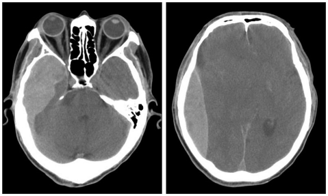
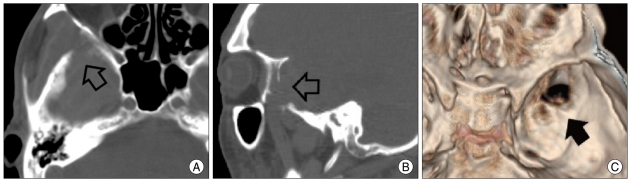
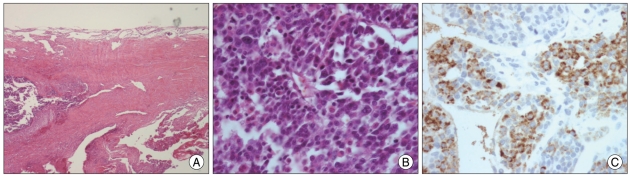
Spontaneous intracranial epidural hematoma (EDH) due to dural metastasis of hepatocellular carcinoma is very rare. A 53-year-old male patient with hepatocellular carcinoma, who was admitted to the department of oncology, was referred to department of neurosurgery because of sudden mental deterioration to semicoma with papillary anisocoria and decerebrate rigidity after transarterial chemoembolization for hepatoma. Brain computed tomography (CT) revealed large amount of acute EDH with severe midline shifting. An emergent craniotomy and evacuation of EDH was performed. Active bleeding from middle cranial fossa floor was identified. There showed osteolytic change on the middle fossa floor with friable mass-like lesion spreading on the overlying dura suggesting metastasis. Pathological examination revealed anaplastic cells with sinusoidal arrangement which probably led to spontaneous hemorrhage and formation of EDH. As a rare cause of spontaneous EDH, dural metastasis from malignancy should be considered.
Keywords: Dural metastasis; Hepatocellular carcinoma; Spontaneous epidural hematoma.
Figures
References
-
- Anegawa S, Hirohata S, Tokutomi T, Kuramoto S. Spontaneous epidural hematoma secondary to dural metastasis from an ovarian carcinoma--case report. Neurol Med Chir (Tokyo) 1989;29:854–856. - PubMed
-
- Aslam E, Imran M, Faridi NM. Bilateral parietal extradural metastatic Ewing's sarcoma simulating acute epidural hematoma. J Coll Physicians Surg Pak. 2006;16:543–544. - PubMed
-
- Cho DY, Liau WR, Chiang IP. Eosinophilic granuloma with acute epidural hematoma : a case report. Pediatr Neurosurg. 2001;35:266–269. - PubMed
-
- Endo M, Hamano M, Watanabe K, Wakai S. [Combined chronic subdural and acute epidural hematoma secondary to metastatic hepatocellular cancer : case report.] No Shinkei Geka. 1999;27:331–334. - PubMed
Publication types
LinkOut - more resources
Full Text Sources
Research Materials
