

Undetermined fibrous tumor with calcification in the cerebellopontine angle
- PMID: 20856670
- PMCID: PMC2941864
- DOI: 10.3340/jkns.2010.48.2.173
Undetermined fibrous tumor with calcification in the cerebellopontine angle
Abstract
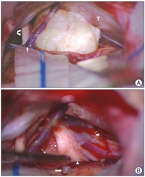
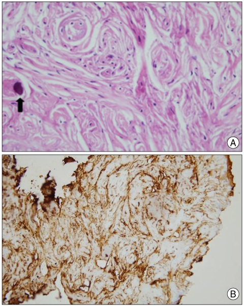
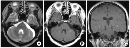
In this report, we introduce an undetermined fibrous tumor with calcification occurring in the cerebellopontine angle (CPA). A 51-year-old woman was admitted with a short history of dizziness. Computed tomography and magnetic resonance images revealed a 2×2×2 cm sized mass at the left CPA which was round and calcified. There was no dura or internal auditory canal involvement. At surgery, the tumor was located at the exit of 7th and 8th cranial nerve complex. It was very firm, bright yellow and well encapsulated. Histologic findings revealed that the tumor was predominantly composed of fibrous component, scant spindle cells and dystrophic calcification. Immunohistochemical staining demonstrated positive for vimentin and negative for epithelial membrane antigen (EMA), S-100 protein, CD34, factor XIIIa and smooth muscle actin. The diagnosis was not compatible with meningioma, schwannoma, metastatic brain tumors, and other fibrous tumors. Although the tumor was resected in total, long term follow-up monitoring is necessary due to the possibility of recurrence.
Keywords: Calcification; Cerebellopontine angle; Immunohistochemistry; Tumor.
Figures

References
-
- Bikmaz K, Cosar M, Kurtkaya-Yapicier O, Iplikcioglu AC, Gokduman CA. Recurrent solitary fibrous tumour in the cerebellopontine angle. J Clin Neurosci. 2005;12:829–832. - PubMed
-
- Brian DM, Caterina G, Michael JL. Ganglioglioma in the cerebellopontine angle in a child. Case report and review of the literature. J Neurosurg. 2007;107:292–296. - PubMed
-
- Dervan PA, Tobin B, O'Connor M. Solitary (localized) fibrous mesothelioma : evidence against mesothelial cell origin. Histopathology. 1986;10:867–875. - PubMed
Publication types
LinkOut - more resources
Full Text Sources
Research Materials
