

A painful glomus tumor on the pulp of the distal phalanx
- PMID: 20856673
- PMCID: PMC2941867
- DOI: 10.3340/jkns.2010.48.2.185
A painful glomus tumor on the pulp of the distal phalanx
Abstract
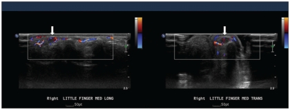
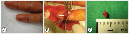
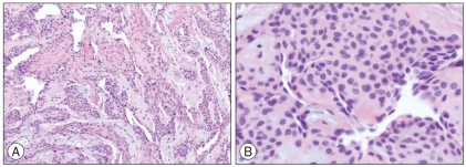
A 52-year-old female patient presented with an 8-year history of progressively intense pain, cold sensitivity, and severe tenderness to palpation of the ulnar side of the tip of her right little finger. Subsequent diagnostic evaluation with ultrasonographic imaging revealed the presence of a glomus tumor in the tender area. Glomus tumors are benign, occurring in the vascular hamartomatous tubercles of the glomus body, which is a myoarterial apparatus typically found in the reticular dermis of the skin. Distal glomus tumors are relatively uncommon, and account for approximately 1% of all hand tumors. Most of them are located in the subungual area because of its high concentration of glomus bodies. We report a case of a glomus tumor with a typical triad of symptoms, yet with a rare location : on the pulp of the ulnar aspect of the distal phalanx of the right little finger.
Keywords: Finger; Glomus tumor; Pain.
Figures
References
-
- Bhaskaranand K, Navadgi BC. Glomus tumour of the hand. J Hand Surg Br. 2002;27:229–231. - PubMed
-
- Carroll RE, Berman AT. Glomus tumors of the hand : review of the literature and report on twenty-eight cases. J Bone Joint Surg Am. 1972;54:691–703. - PubMed
-
- Fornage BD. Glomus tumors in the fingers : diagnosis with US. Radiology. 1988;167:183–185. - PubMed
-
- Heim U, Hänggi W. Subungual glomus tumors. Value of the direct dorsal approach. Ann Chir Main. 1985;4:51–54. - PubMed
-
- Kale SS, Rao VK, Bentz ML. Glomus tumor of the index finger. J Craniofac Surg. 2006;17:801–804. - PubMed
Publication types
LinkOut - more resources
Full Text Sources
