. 2010 Sep 15;5(9):e12772.
doi: 10.1371/journal.pone.0012772.

Induction of ER stress in macrophages of tuberculosis granulomas

Affiliations

Induction of ER stress in macrophages of tuberculosis granulomas

Tracie A Seimon et al. PLoS One. .

Abstract

Background: The endoplasmic reticulum (ER) stress pathway known as the Unfolded Protein Response (UPR) is an adaptive survival pathway that protects cells from the buildup of misfolded proteins, but under certain circumstances it can lead to apoptosis. ER stress has been causally associated with macrophage apoptosis in advanced atherosclerosis of mice and humans. Because atherosclerosis shares certain features with tuberculosis (TB) with regard to lesional macrophage accumulation, foam cell formation, and apoptosis, we investigated if the ER stress pathway is activated during TB infection.

Principal findings: Here we show that ER stress markers such as C/EBP homologous protein (CHOP; also known as GADD153), phosphorylated inositol-requiring enzyme 1 alpha (Ire1α) and eukaryotic initiation factor 2 alpha (eIF2α), and activating transcription factor 3 (ATF3) are expressed in macrophage-rich areas of granulomas in lungs of mice infected with virulent Mycobacterium tuberculosis (Mtb). These areas were also positive for numerous apoptotic cells as assayed by TUNEL. Microarray analysis of human caseous TB granulomas isolated by laser capture microdissection reveal that 73% of genes involved in the UPR are upregulated at the mRNA transcript level. The expression of two ER stress markers, ATF3 and CHOP, were also increased in macrophages of human TB granulomas when assayed by immunohistochemistry. CHOP has been causally associated with ER stress-induced macrophage apoptosis. We found that apoptosis was more abundant in granulomas as compared to non-granulomatous tissue isolated from patients with pulmonary TB, and apoptosis correlated with CHOP expression in areas surrounding the centralized areas of caseation.

Conclusions: In summary, ER stress is induced in macrophages of TB granulomas in areas where apoptotic cells accumulate in mice and humans. Although macrophage apoptosis is generally thought to be beneficial in initially protecting the host from Mtb infection, death of infected macrophages in advanced granulomas might favor dissemination of the bacteria. Therefore future work is needed to determine if ER-stress is causative for apoptosis and plays a role in the host response to infection.


Conflict of interest statement

Competing Interests: The authors have declared that no competing interests exist.

Figures

Figure 1
Figure 1. Apoptotic macrophages in CHOP-positive regions of Mtb-infected mouse lung.
Mice were infected by aerosol with Mtb. Eight weeks later lungs were fixed and embedded in paraffin. Granulomas are identified in the center of each image and have rings of lymphocytes (dark blue) surrounding a central macrophage-rich core. A and B. Immunohistochemistry was performed using an antibody against CHOP and a normal IgG control. Slides were counter-stained with hematoxylin. A. Low power image of CHOP staining (brown) in several granulomas present (4x magnification, red arrows). B. IgG and CHOP staining (10x magnification, bar represents 1 mm). C. A serial section was stained for macrophages using an anti-Mac-3 antibody (green), and for apoptotic cells by TUNEL (red). Nuclei were stained using DAPI. Left and middle panels (20x and 40x magnification respectively) are merged images of Mac3, TUNEL, and DAPI stain. Black and white dashed boxes indicate corresponding areas of the granuloma of adjacent sections. Red arrows indicate TUNEL positive cells that were also positive for Mac3 and DAPI. Far right panel is the same area in a serial section, also shown in A and B, stained for CHOP by immunohistochemistry (40x magnification, bar represents 100 µm).
Figure 2
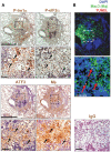
Figure 2. UPR markers are induced in macrophages residing in granulomas during Mtb infection in mice.
Mice were infected as described in Figure 1. A and B. Immunohistochemistry was performed on lung sections using an antibody against Ser724 Ire1α (P- Ire1α), Ser51 eIF2α (P- eIF2α), ATF3, the macrophage (Mφ) marker AIA31240, and normal rabbit IgG. Slides were counter-stained with hematoxylin (upper panels, magnification 10x, bar represents 1 mm; lower panels, magnification 40x, bar represents 100 µm). B. A serial section was stained for macrophages using an anti-Mac-3 antibody (green), and for apoptotic cells by TUNEL (red). Nuclei were stained using DAPI (bar represents 100 µm). Dashed boxes in A and B indicate similar areas of the granuloma in adjacent sections that were enlarged in the corresponding images for clarity. The black arrows represent similar areas in the serial sections that are positive for all four markers. Red arrows indicate cells that were positive for TUNEL and the macrophage marker Mac-3.
Figure 3
Figure 3. ER stress-induced genes are upregulated in human TB granulomas.
RNA isolated from caseous granulomas by laser capture microdissection from 3 TB patients was subjected to microarray analysis. All genes in the database were ranked and a hierarchical list of each gene in relative transcript abundance was created. Shown is a comparison of ER resident genes that that have not been shown to be regulated by ER stress, common innate immune receptors such as scavenger receptors, TLRs, and macrophage markers, and genes known to participate in the Unfolded Protein Response or ER stress pathway. Error bars represent the standard deviation of caseous granulomas from three independent patients compared in the microarray. Genes represented by a red bars had a ranking above 10 and were represented in the top third of all genes in relative transcript abundance, and the genes represented by green bars fell below that threshold.
Figure 4
Figure 4. Induction of ER stress markers in human caseous TB granulomas.
A and B. Lung sections from TB patients containing granulomas were stained by immunohistochemistry using an antibody against CHOP, normal IgG control, ATF3, and the macrophage marker CD68. Slides were counter-stained with hematoxylin. A. CHOP staining (brown), but not the IgG control, is seen around the central area of caseation (bar represents 100 µm). B. Low and high power magnification shows CHOP staining in areas that are also positive for ATF3 and CD68 (hatched box). The bar in the upper panel represents 1 mm while the bar in the lower panel represents 100 µm. Also shown are multinucleated giant cells surrounding the granuloma also positive for CHOP, ATF3, and CD68 (red arrows).
Figure 5
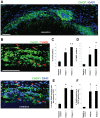
Figure 5. Increased apoptosis in CHOP-positive regions of human TB granulomas.
Lung sections from TB patients containing granulomas were stained for CHOP (green), TUNEL (red), and nuclei (DAPI; blue). Patients 1 and 2 were positive for granulomas and the negative control was uninvolved lung parenchyma from a TB patient. A. CHOP staining (green) is seen around the central area of caseation (bar represents 100 µm). B. High power magnification shows TUNEL staining in areas that are also positive for CHOP and the DNA stain DAPI (red arrows, bar represents 100 µm). C. Quantification of the percent of CHOP-positive cells from 4 random fields of view. The number of CHOP-positive cells were expressed as a percent of total DAPI-positive cells in each field (n = 4 fields/patient). D. Quantification of the percent of TUNEL-positive cells from 4 random fields of view. The number of cells positive for both TUNEL and DAPI were expressed as a percent of total DAPI-positive cells in each field (n = 4 fields/patient). E. Quantification of the percent of total TUNEL-positive cells that express CHOP. The number of cells positive for TUNEL, DAPI, and CHOP were expressed as a percent of total number of TUNEL-positive cells in each field (n = 4 fields/patient). F. Quantification of the percent TUNEL-positivity in the CHOP expressing population. The number of cells positive for TUNEL, DAPI, and CHOP were expressed as a percent of total number of CHOP-positive cells in each field (n = 4 fields/patient). Differences between values with symbols and no symbols, or between values with different symbols, are statistically significant by ANOVA followed by post-hoc Student-Newman-Keuls test (P<0.05).
Figure 6
Figure 6. BCG-induced macrophage death is enhanced by thapsigargin.
A. Murine peritoneal macrophages were infected with the indicated MOI of BCG in the presence or absence of 0.5 µM thapsigargin (an ER stressor). After 24 hours the macrophages were analyzed for apoptosis, and the data are expressed as the percent of total cells that stained with annexin V and propidium iodide (mean ± SEM; n = 4). Representative fluorescent images are shown. The presence of symbols or differing symbols indicate P<0.05; identical symbols or the absence of symbols indicate differences that are not significant by ANOVA post-hoc Student-Newman-Keuls test.

Similar articles

Cited by

References

    1. Kim I, Xu W, Reed JC. Cell death and endoplasmic reticulum stress: disease relevance and therapeutic opportunities. Nat Rev Drug Discov. 2008;7:1013–1030. - PubMed
    1. Medigeshi GR, Lancaster AM, Hirsch AJ, Briese T, Lipkin WI, et al. West Nile virus infection activates the unfolded protein response, leading to CHOP induction and apoptosis. The Journal of Virology. 2007;81:10849–10860. - PMC - PubMed
    1. Williams BL, Lipkin WI. Endoplasmic reticulum stress and neurodegeneration in rats neonatally infected with Borna Disease virus. The Journal of Virology. 2006;80:8613–8626. - PMC - PubMed
    1. Malhotra J, Kaufman R. Endoplasmic reticulum stress and oxidative stress: A vicious cycle or double edged sword. Antioxidants and Redox Signaling. 2007;9:2277–2294. - PubMed
    1. Tamaki N, Hatano E, Taura K, Tada M, Kodama Y, et al. CHOP deficiency attenuates cholestasis-induced liver fibrosis by reduction of hepatocyte injury. Am J Physiol Gastrointest Liver Physiol. 2008;294:G498–G505. - PubMed

Publication types

MeSH terms

Substances