Novel photosensitizers trigger rapid death of malignant human cells and rodent tumor transplants via lipid photodamage and membrane permeabilization
- PMID: 20856679
- PMCID: PMC2939899
- DOI: 10.1371/journal.pone.0012717
Novel photosensitizers trigger rapid death of malignant human cells and rodent tumor transplants via lipid photodamage and membrane permeabilization
Abstract
Background: Apoptotic cascades may frequently be impaired in tumor cells; therefore, the approaches to circumvent these obstacles emerge as important therapeutic modalities.
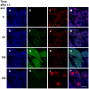
Methodology/principal findings: Our novel derivatives of chlorin e(6), that is, its amide (compound 2) and boronated amide (compound 5) evoked no dark toxicity and demonstrated a significantly higher photosensitizing efficacy than chlorin e(6) against transplanted aggressive tumors such as B16 melanoma and M-1 sarcoma. Compound 5 showed superior therapeutic potency. Illumination with red light of mammalian tumor cells loaded with 0.1 µM of 5 caused rapid (within the initial minutes) necrosis as determined by propidium iodide staining. The laser confocal microscopy-assisted analysis of cell death revealed the following order of events: prior to illumination, 5 accumulated in Golgi cysternae, endoplasmic reticulum and in some (but not all) lysosomes. In response to light, the reactive oxygen species burst was concomitant with the drop of mitochondrial transmembrane electric potential, the dramatic changes of mitochondrial shape and the loss of integrity of mitochondria and lysosomes. Within 3-4 min post illumination, the plasma membrane became permeable for propidium iodide. Compounds 2 and 5 were one order of magnitude more potent than chlorin e(6) in photodamage of artificial liposomes monitored in a dye release assay. The latter effect depended on the content of non-saturated lipids; in liposomes consisting of saturated lipids no photodamage was detectable. The increased therapeutic efficacy of 5 compared with 2 was attributed to a striking difference in the ability of these photosensitizers to permeate through hydrophobic membrane interior as evidenced by measurements of voltage jump-induced relaxation of transmembrane current on planar lipid bilayers.
Conclusions/significance: The multimembrane photodestruction and cell necrosis induced by photoactivation of 2 and 5 are directly associated with membrane permeabilization caused by lipid photodamage.
Conflict of interest statement
Figures


Similar articles
-
Photolon™ --photosensitization induces apoptosis via ROS-mediated cross-talk between mitochondria and lysosomes.Int J Oncol. 2011 Oct;39(4):821-31. doi: 10.3892/ijo.2011.1109. Epub 2011 Jul 1. Int J Oncol. 2011. PMID: 21725591
-
Mitochondrial photodamage and PDT-induced apoptosis.J Photochem Photobiol B. 1998 Feb;42(2):89-95. doi: 10.1016/s1011-1344(97)00127-9. J Photochem Photobiol B. 1998. PMID: 9540214
-
Cell death via mitochondrial apoptotic pathway due to activation of Bax by lysosomal photodamage.Free Radic Biol Med. 2011 Jul 1;51(1):53-68. doi: 10.1016/j.freeradbiomed.2011.03.042. Epub 2011 Apr 8. Free Radic Biol Med. 2011. PMID: 21530645
-
Cell death and growth arrest in response to photodynamic therapy with membrane-bound photosensitizers.Biochem Pharmacol. 2003 Oct 15;66(8):1651-9. doi: 10.1016/s0006-2952(03)00539-2. Biochem Pharmacol. 2003. PMID: 14555246 Review.
-
Recent Advances in Activatable Organic Photosensitizers for Specific Photodynamic Therapy.Chempluschem. 2020 May;85(5):948-957. doi: 10.1002/cplu.202000203. Chempluschem. 2020. PMID: 32401421 Review.
Cited by
-
Nanostructural and nanomechanical alterations of photosensitized lipid membranes due to light induced formation of reactive oxygen species.Sci Rep. 2025 Jan 2;15(1):110. doi: 10.1038/s41598-024-83758-w. Sci Rep. 2025. PMID: 39747172 Free PMC article.
-
Curvature-Assisted Vesicle Explosion Under Light-Induced Asymmetric Oxidation.Adv Sci (Weinh). 2024 Oct;11(38):e2400504. doi: 10.1002/advs.202400504. Epub 2024 Aug 13. Adv Sci (Weinh). 2024. PMID: 39136143 Free PMC article.
-
Effect of ricin on photodynamic damage to the plasma membrane.Dokl Biochem Biophys. 2013 Mar-Apr;449:84-6. doi: 10.1134/S1607672913020087. Epub 2013 May 9. Dokl Biochem Biophys. 2013. PMID: 23657653 No abstract available.
-
Comparison of light-induced formation of reactive oxygen species and the membrane destruction of two mesoporphyrin derivatives in liposomes.Sci Rep. 2019 Aug 5;9(1):11312. doi: 10.1038/s41598-019-47841-x. Sci Rep. 2019. PMID: 31383921 Free PMC article.
-
Perfluorocarbon Nanoemulsions with Fluorous Chlorin-Type Photosensitizers for Antitumor Photodynamic Therapy in Hypoxia.Int J Mol Sci. 2023 Apr 28;24(9):7995. doi: 10.3390/ijms24097995. Int J Mol Sci. 2023. PMID: 37175700 Free PMC article.
References
-
- O'Connor AE, Gallagher WM, Byrne AT. Porphyrin and nonporphyrin photosensitizers in oncology: preclinical and clinical advances in photodynamic therapy. Photochem Photobiol. 2009;85:1053–1074. - PubMed
-
- Maisch T. A new strategy to destroy antibiotic resistant microorganisms: antimicrobial photodynamic treatment. Mini Rev Med Chem. 2009;9:974–983. - PubMed
-
- Robertson CA, Evans DH, Abrahamse H. Photodynamic therapy (PDT): a short review on cellular mechanisms and cancer research applications for PDT. J Photochem Photobiol B. 2009;96:1–8. - PubMed
-
- Koudinova NV, Pinthus JH, Brandis A, Brenner O, Bendel P, et al. Photodynamic therapy with Pd-bacteriopheophorbide (TOOKAD): successful in vivo treatment of human prostatic small cell carcinoma xenografts. Int J Cancer. 2003;104:782–789. - PubMed
-
- Brandis A, Mazor O, Neumark E, Rosenbach-Belkin V, Salomon Y, Scherz A. Novel water-soluble bacteriochlorophyll derivatives for vascular-targeted photodynamic therapy: synthesis, solubility, phototoxicity and the effect of serum proteins. Photochem Photobiol. 2005;81:983–993. - PubMed
Publication types
MeSH terms
Substances
LinkOut - more resources
Full Text Sources

