. 2010 Sep 15;5(9):e12753.
doi: 10.1371/journal.pone.0012753.

Loss of STOP protein impairs peripheral olfactory neurogenesis

Affiliations

Loss of STOP protein impairs peripheral olfactory neurogenesis

Karelle Benardais et al. PLoS One. .

Abstract

Background: STOP (Stable Tubulin-Only Polypeptide) null mice show behavioral deficits, impaired synaptic plasticity, decrease in synaptic vesicular pools and disturbances in dopaminergic transmission, and are considered a neurodevelopmental model of schizophrenia. Olfactory neurons highly express STOP protein and are continually generated throughout life. Experimentally-induced loss of olfactory neurons leads to epithelial regeneration within two months, providing a useful model to evaluate the role played by STOP protein in adult olfactory neurogenesis.

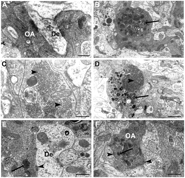
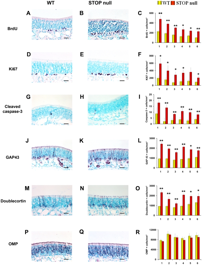
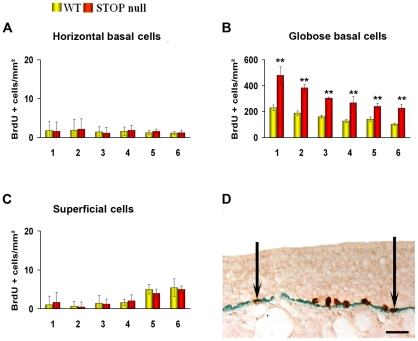
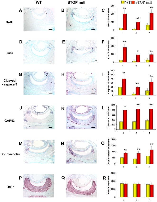
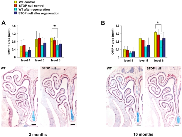
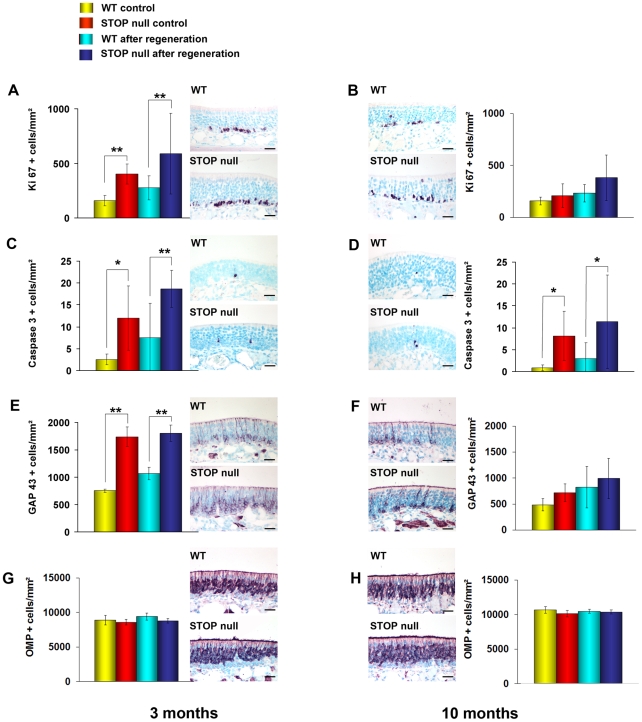
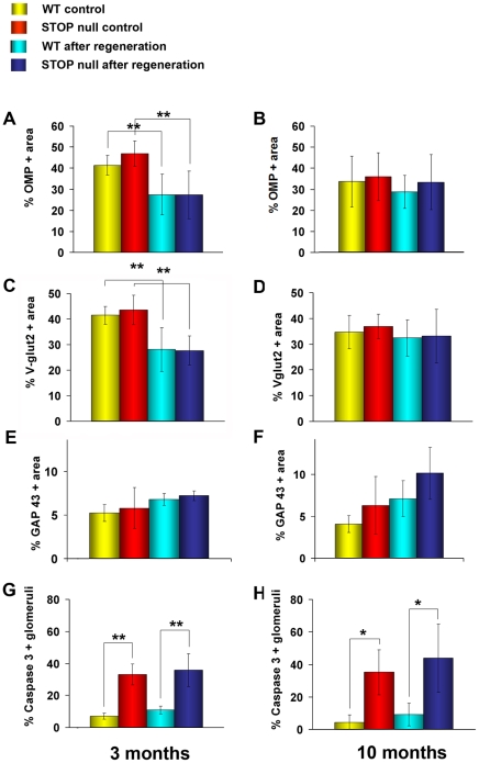
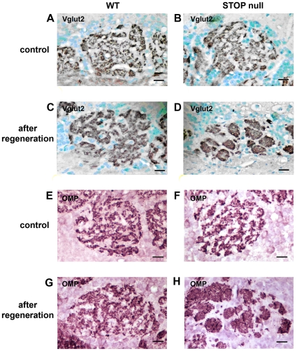
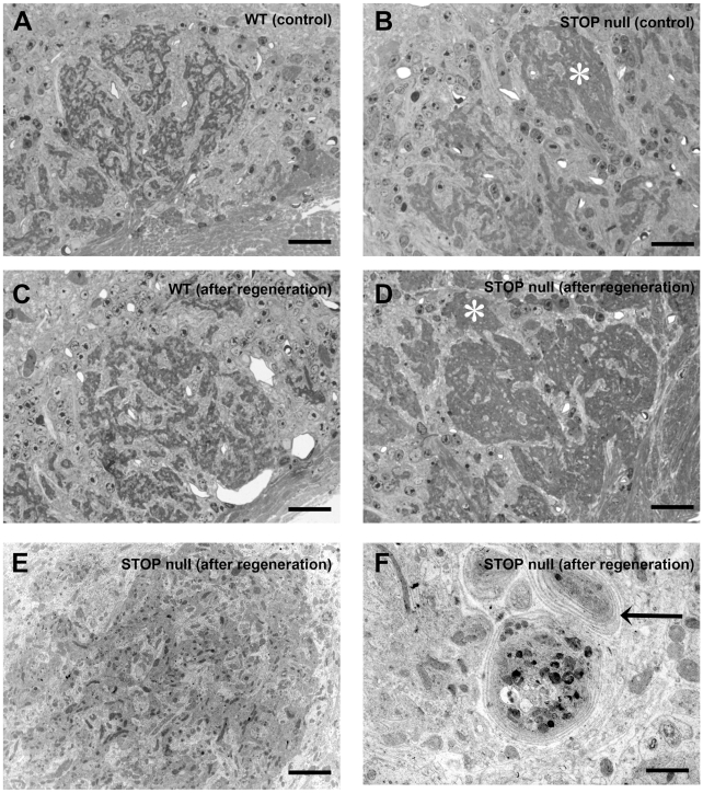
Methodology/principal findings: Immunocytochemistry and electron microscopy were used to study the structure of the glomerulus in the main olfactory bulb and neurogenesis in the neurosensorial epithelia. In STOP null mice, olfactory neurons showed presynaptic swellings with tubulovesicular profiles and autophagic-like structures. In olfactory and vomeronasal epithelia, there was an increase in neurons turnover, as shown by the increase in number of proliferating, apoptotic and immature cells with no changes in the number of mature neurons. Similar alterations in peripheral olfactory neurogenesis have been previously described in schizophrenia patients. In STOP null mice, regeneration of the olfactory epithelium did not modify these anomalies; moreover, regeneration resulted in abnormal organisation of olfactory terminals within the olfactory glomeruli in STOP null mice.

Conclusions/significance: In conclusion, STOP protein seems to be involved in the establishment of synapses in the olfactory glomerulus. Our results indicate that the olfactory system of STOP null mice is a well-suited experimental model (1) for the study of the mechanism of action of STOP protein in synaptic function/plasticity and (2) for pathophysiological studies of the mechanisms of altered neuronal connections in schizophrenia.


Conflict of interest statement

Competing Interests: The authors have declared that no competing interests exist.

Figures

Figure 1
Figure 1. Ultrastructure of olfactory bulb glomeruli.
Electron microscopy micrographs of olfactory bulb glomeruli in WT (A) and STOP null (B–F) mice at 3 to 6 months of age. In STOP null mice, olfactory axon endings are filled with autophagic-like structures (B, arrow), tubulovesicular profiles (C, arrowhead), or both (D). When few autophagic structures were present (arrows) (E, F), olfactory axons endings could be identified by the presence of synaptic vesicles and postsynaptic densities (arrowheads) (E, F). De: dentrite; OA: olfactory axon. Scale bar: 0,5 µm (A, C, E, F); 1 µm (B, D).
Figure 2
Figure 2. Neurogenesis in the olfactory epithelium.
Localisation and mean density of BrdU (A–C), Ki67 (D–F), cleaved caspase 3 (G–I), GAP 43 (J–L), doublecortin (M–O) and OMP (P–R) labelled cells in the olfactory epithelium of WT and STOP null mice. The x-axis refers to the six levels studied, from rostral (level 1) to caudal (level 6). A statistically significant increase in the number of proliferating (BrdU and Ki67 positive cells), apoptotic (caspase 3 positive cells) and immature neurons (GAP 43 and doublecortin positive cells) was observed in STOP null mice as compared to WT mice. There was no difference in the number of mature OMP expressing neurons between the two genotypes. All values are represented as mean +/− SEM, *p<0.05, **p<0.01. Scale bar: 25 µm.
Figure 3
Figure 3. Localisation of BrdU-labelled cells in the olfactory epithelium.
Mean density of BrdU-labelled cells in cytokeratine 5 positive HBC layer (A), cytokeratine 5 negative GBC layer (B) and superficial layer (C) in the olfactory epithelium of WT and STOP null mice. The x-axis refers to the six levels studied, from rostral (level 1) to caudal (level 6). Double immunolabelling (arrows) for BrdU (brown) and cytokeratin 5 (green) in the olfactory epithelium of a STOP null mouse is illustrated in D. An increase in globose basal cells but not in horizontal basal and superficial cells was observed in STOP null mice as compared to WT mice. All values are represented as mean +/− SEM, **p<0.01. Scale bar: 30 µm.
Figure 4
Figure 4. Neurogenesis in the vomeronasal epithelium.
Localisation and mean density of BrdU (A–C), Ki67 (D–F), cleaved caspase 3 (G–I), GAP 43 (J–L), doublecortin (M–O) and OMP (P–R) labelled cells in the vomeronasal epithelium of WT and STOP null mice. The x-axis refers to the three levels studied, from rostral (level 1) to caudal (level 3), where the vomeronasal organ was present. A statistically significant increase in proliferating (BrdU and Ki67 positive cells), apoptotic (caspase 3 positive cells) and immature neurons (GAP 43 and doublecortin positive cells), but not mature OMP positive neurons was observed in STOP null mice as compared to WT mice. All values are represented as mean +/− SEM, *p<0.05, **p<0.01. Scale bar: 100 µm.
Figure 5
Figure 5. Regeneration of the olfactory epithelium.
Mean epithelial OMP positive area before and after regeneration at the three levels 4, 5, 6 in WT and STOP null mice at two different ages. The x-axis refers to the three levels studied, from rostral (level 4) to caudal (level 6), where turbinates are most developed and olfactory epithelium most abundant. There is no difference in the ability of olfactory epithelium to regenerate at the three levels studied between WT and STOP null mice in 3 month-old (A) and 10 month-old (B) animals. All values are represented as mean +/− SEM, *p<0.05. The photomicrographs in A and B illustrate OMP immunostaining in the olfactory epithelium of animals after regeneration. Scale bar: 500 µm.
Figure 6
Figure 6. Neurogenesis in the olfactory epithelium after regeneration.
Mean density of Ki 67 (A, B), caspase 3 (C, D), GAP 43 (E, F) and OMP (G, H) positive cells in the olfactory epithelium of WT and STOP null mice at two different ages. In the 3-month-old groups (A, C, E, G), apoptotic, proliferating and immature, but not mature neurons are more numerous in STOP null mice as compared to WT mice, both in control animals and after regeneration. In the 10-month-old groups (B, D, F, H) only the number of caspase 3 positive neurons (D) was increased in STOP null mice as compared to WT mice, in controls and after regeneration. All values are represented as mean +/− SEM, *p<0.05, **p<0.01. The photomicrographs illustrate immunostaining in animals after regeneration. Scale bar: 25 µm.
Figure 7
Figure 7. Formation of glomeruli after regeneration.
Mean percentage of OMP (A, B), Vglut2 (C, D), GAP 43 (E, F) positive glomerular areas and mean percentage of caspase 3 (G, H) positive glomeruli in WT and STOP null mice at two different ages. In control groups and after regeneration there was no difference between the two genotypes concerning OMP, Vglut2 and GAP 43 immunolabelling at both ages. In controls and after regeneration, the number of glomeruli with apoptotic fibers was greater in STOP null mice as compared to WT mice at both ages. All values are represented as mean +/− SEM, *p<0.05, **p<0.01. The photomicrographs illustrate immunostaining in animals after regeneration. Scale bar: 25 µm.
Figure 8
Figure 8. Glomerular structure after regeneration in 10-month-old mice.
Localisation of Vglut2 (A–D) and OMP (E–H) immunolabelling in olfactory bulb glomeruli of WT (A, C, E, G) and STOP null (B, D, F H) mice in controls (A, B, E, F) and after regeneration (C, D, G, H). Note the characteristic feature of a mosaicism between Vglut2 or OMP positive fibers and Vglut2 or OMP negative dentrites in WT mice either in controls or after regeneration (A, E and C, G respectively). In STOP null mice, a clumped aspect of either Vglut2 (D) or OMP (H) positive fibers was observed. Scale bar: 20 µm.
Figure 9
Figure 9. Glomeruli ultrastructure after regeneration in 10-month-old mice.
Semithin sections (A–D) and electron microscopy micrographs (E, F) of olfactory bulb glomeruli in WT (A, C) and STOP null (B, D, E, F) mice. In STOP null controls, some olfactory endings are densely packed (*) (B); after regeneration this aspect is more pronounced (D). E illustrates the ultrastructure of a densely packed terminal area in D (*); olfactory axons are densely packed and associated with few electron clear dendrites area. F illustrates concentrically organized axon terminals (F, arrow) in STOP null mice following regeneration. Scale bar: 20 µm (A–D); 5 µm (E); 3 µm (F).

Similar articles

Cited by

References

    1. Bosc C, Andrieux A, Job D. Stop proteins. Biochemistry. 2003;42:12125–12132. - PubMed
    1. Job D, Fischer EH, Margolis RL. Rapid disassembly of cold-stable microtubules by calmodulin. Proc Natl Acad Sci USA. 1981;78:4679–4682. - PMC - PubMed
    1. Guillaud L, Bosc C, Fourest-Lieuvin A, Denarier E, Pirollet F, et al. STOP proteins are responsible for the high degree of microtubule stabilization observed in neuronal cells. J Cell Biol. 1998;142:167–179. - PMC - PubMed
    1. Andrieux A, Salin PA, Vernet M, Kujala P, Baratier J, et al. The suppression of brain cold-stable microtubules in mice induces synaptic defects associated with neuroleptic-sensitive behavioural disorders. Genes Dev. 2002;16:2350–2364. - PMC - PubMed
    1. Brun P, Begou M, Andrieux A, Mouly-Badina L, Clerget M. Dopaminergic transmission in STOP null mice. J Neurochem. 2005;94:63–73. - PubMed

Publication types

Substances

LinkOut - more resources