. 2010 Sep 15;5(9):e12765.
doi: 10.1371/journal.pone.0012765.

Mammalian Otolin: a multimeric glycoprotein specific to the inner ear that interacts with otoconial matrix protein Otoconin-90 and Cerebellin-1

Affiliations

Mammalian Otolin: a multimeric glycoprotein specific to the inner ear that interacts with otoconial matrix protein Otoconin-90 and Cerebellin-1

Michael R Deans et al. PLoS One. .

Abstract

Background: The mammalian otoconial membrane is a dense extracellular matrix containing bio-mineralized otoconia. This structure provides the mechanical stimulus necessary for hair cells of the vestibular maculae to respond to linear accelerations and gravity. In teleosts, Otolin is required for the proper anchoring of otolith crystals to the sensory maculae. Otoconia detachment and subsequent entrapment in the semicircular canals can result in benign paroxysmal positional vertigo (BPPV), a common form of vertigo for which the molecular basis is unknown. Several cDNAs encoding protein components of the mammalian otoconia and otoconial membrane have recently been identified, and mutations in these genes result in abnormal otoconia formation and balance deficits.

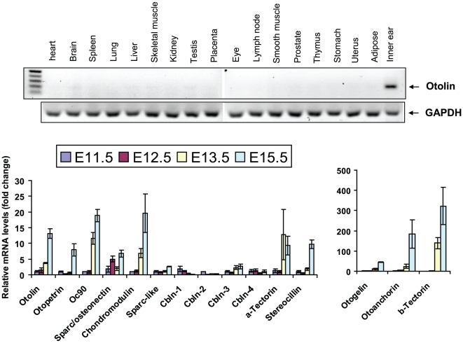
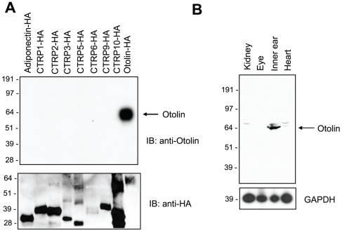
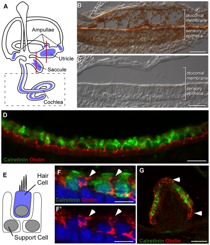
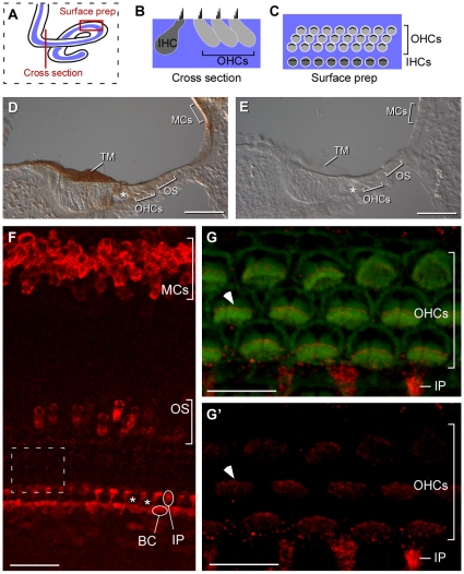
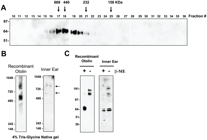
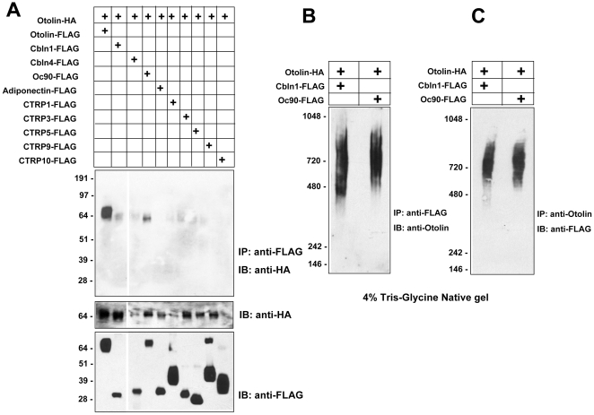
Principal findings: Here we describe the cloning and characterization of mammalian Otolin, a protein constituent of otoconia and the otoconial membrane. Otolin is a secreted glycoprotein of ∼70 kDa, with a C-terminal globular domain that is homologous to the immune complement C1q, and contains extensive posttranslational modifications including hydroxylated prolines and glycosylated lysines. Like all C1q/TNF family members, Otolin multimerizes into higher order oligomeric complexes. The expression of otolin mRNA is restricted to the inner ear, and immunohistochemical analysis identified Otolin protein in support cells of the vestibular maculae and semi-circular canal cristae. Additionally, Otolin forms protein complexes with Cerebellin-1 and Otoconin-90, two protein constituents of the otoconia, when expressed in vitro. Otolin was also found in subsets of support cells and non-sensory cells of the cochlea, suggesting that Otolin is also a component of the tectorial membrane.

Conclusion: Given the importance of Otolin in lower organisms, the molecular cloning and biochemical characterization of the mammalian Otolin protein may lead to a better understanding of otoconial development and vestibular dysfunction.


Conflict of interest statement

Competing Interests: The authors have declared that no competing interests exist.

Figures

Figure 1
Figure 1. Cloning of the mouse otolin cDNA.
A, The deduced mouse and human otolin proteins consist of four domains— a signal peptide (S), an N-terminal region with four Cys residues, a collagen domain with 75 Gly-X-Y repeats, and a globular C1q domain. All Cys residues with their amino acid positions are indicated with a ball-and-stick. Note that human otolin has three more Cys residues located in the collagen domain compared to mouse otolin. B, The exon/intron structures of human and mouse otolin gene. Dashed boxes indicate 5′ and 3′UTR that cannot be determined due to lack of homology between the human and mouse gene. The size of each exon and intron are indicated. The region of the cDNA encoded by each exon is indicated by dash line.
Figure 2
Figure 2. Alignment of otolin sequences from multiple species.
ClustalW alignment of Otolin protein sequences extracted from the draft genome sequences of human (Homo sapiens; accession number NP_001073909), mouse (Mus musculus; DQ002405), cow (Bos taurus; XP_603387.3), opossum (Monodelphis domestica; XP_001369147), chicken (Gallus gallus; XP_426716.2), platypus (Ornithorhynchus anatinus; XP_001512453), and zebrafish (Danio rerio; NP_001093211). Identical amino acids are shaded and gaps are indicated by a dash line. All the conserved Cys residues are indicated by a green ball-and-stick. Signal peptide (yellow line), N-terminus (purple line), collagen domain (blue line), and globular C1q domain (red line) are indicated. Conserved residues found in all C1q/TNF family members are indicated by arrows.
Figure 3
Figure 3. Expression of otolin transcripts in mouse tissues.
A, The expression profile of otolin mRNA in various mouse tissues as revealed by a semi-quantitative PCR. B, Real time PCR analysis of the expression profiles of otolin and other known inner ear genes during development. All expression data are normalized to 18S RNA.
Figure 4
Figure 4. Specificity of Otolin Antibody.
A, Western blot analysis of the HA-tagged adiponectin, CTRP1, CTRP2, CTRP3, CTRP5, CTRP6, CTRP9, CTRP10, and otolin proteins using an anti-otolin (top panel) or an anti-HA antibody (bottom panel). B, Western blot analysis of mouse kidney, eye, inner ear, and heart tissue lysates using the anti-otolin antibody.
Figure 5
Figure 5. Distribution of Otolin protein within the vestibular sensory epithelia of the inner ear.
A, Hair cell receptors are located in one of six sensory epithelia as indicated by blue shading in this illustration of the mouse inner ear. Vestibular hair cells are found in the maculae of the utricle and saccule and the cristae of the semi-circular canal ampullae. Auditory hair cells are found exclusively in the cochlea. Red lines indicate the relative position of the tissue sections illustrated in the remaining panels. Dashed box indicates the auditory region described in greater detail in Fig. 6B , Histochemical detection of Otolin protein in P2 utricle. Otolin is present in the extracellular matrix of the otoconial membrane and within cells of the sensory epithelia. C, No signal is present in control experiments lacking Otolin antisera. D, Immunofluorescent labeling of Otolin (red) and the hair cell marker Calretinin (green) reveals little colocalization in the P2 saccule, indicating that Otolin is synthesized by support cells. Note that the otoconial membrane is not preserved during this labeling protocol. E, Vestibular hair cells and support cells form a pseudostratified epithelium, with the support cell nuclei located below the hair cells and support cell processes surrounding individual hair cells. F&F', High power confocal image demonstrating Otolin immunofluorescence (red, arrowheads) in support cell processes located between neighboring hair cells (green, F). G, In the semi-circular canal cristae, Otolin is expressed in support cells surrounding the hair cells (green) and located adjacent to the hair cell regions (arrowheads). Scale bars: 50 µm B,C,G; 25 µm D, 10 µm F,F'.
Figure 6
Figure 6. Distribution of Otolin protein in the cochlea.
A–C, The distribution of Otolin in the auditory system was evaluated in cross sections cut through the cochlea (as indicated by a red line) or whole mount surface preparations (outlined by a red box). (B) In cross section the profile of cochlear cell types and overlying tectorial membrane are visible. (C) Surface preparations provide a view of the apical surface of cochlear cells without the tectorial membrane. The relative position and organization of cochlear cells in these two views are illustrated using inner hair cells (IHCs) and outer hair cells (OHCs) for reference. (D) Histochemical detection of Otolin in neonatal (P2) mouse cross sections reveals Otolin protein throughout the tectorial membrane that contacts auditory hair cells. A subset of cochlear support cells is labeled surrounding the IHCs (asterisk) in addition to marginal cells and the outer sulcus. (E), No signal is present in control experiments lacking Otolin antisera. (F) In surface preparations prepared from E18.5 mouse, Otolin (red) can be visualized in inner phalangeal cells (IP) and border cells (BC), two classes of support cells located adjacent to the IHCs. The positions of individual examples are indicated. Although the IHCs are not labeled in this image the position of two IHCs are marked by asterisks. Additional non-sensory cells producing Otolin that contribute to formation of the tectorial membrane can also be visualized in cochlear whole mounts. This includes marginal cells of the stria vascularis and a small population of cells in the outer sulcus. (G), High magnification confocal imaging of OHC stereocilia labeled with phalloidin (green) also shows Otolin protein (red) at the tips of the stereocilia bundle (one example marked by arrowhead) resulting from contact between the stereocilia and the Otolin-rich tectorial membrane. (G') Otolin immunofluorescence detected in the red channel from panel G. The region being imaged is indicated by the dashed box in F. TM (Tectorial Membrane), MCs (Marginal Cells), IHC (Inner Hair Cell), OHC (Outer Hair Cell), IP (Inter Phalangeal Cell), BC (Border Cell), OS (Outer Sulcus). Scale bars: 100 µm D,E; 20 µm F, 10 µm G,G'.
Figure 7
Figure 7. Otolin is a secreted glycoprotein with extensive posttranslational modifications.
A, Western blot analysis of the cell pellets (P) and supernatant (S) from transfected HEK293T cells using the anti-FLAG antibody. B, Recombinant otolin-FLAG was incubated with (+) or without (−) peptide, N-glycosidase F (PNGaseF), to determine the presence of N-linked glycans. Proteins were immunoblotted with the anti-FLAG antibody. C, Recombinant otolin-FLAG was incubated with (+) or without (−) chondroitinase ABC or O-glycosidase to determine the presence of chondroitin sulfate proteoglycans and O-linked glycans, respectively. Chondroitinase ABC preferentially digests hyaluronic acid at pH 6.8 and chondroitin sulfate at pH 8. Proteins were immunoblotted with the anti-FLAG antibody. D, A metaperiodate oxidation-based method was used to detect the presence of carbohydrate moieties on recombinant otolin (See Materials and Methods ). E, Mass spectrometry analyses of recombinant otolin. All lysine residues that lie within the consensus sequence [GXKG(E/D)] are highlighted in blue, and those that are glycosylated are indicated with a square-and-stick. All proline residues (in the Gly-X-Pro context) that lie within the collagen domain are highlighted in red, and those that are hydroxylated are indicated with a ball-and-stick.
Figure 8
Figure 8. Otolin assembles into higher order multimeric complexes.
A, Supernatant containing FLAG-tagged otolin was loaded onto superdex 200 FPLC column and 0.5 ml fractions were collected. Fractions 10 to 27 were analyzed by immunoblot analysis using the anti-FLAG antibody. Arrows with the molecular weight markers of 669, 440, 232, and 158 kDa correspond to the peak elution fraction of molecular standard thyroglobulin, ferritin, catalase, and aldolase, respectively. B, 4% Tris-Glycine native gel immunoblot analysis [see MATERIALS AND METHODS ] of purified recombinant otolin and endogenous otolin (indicated by arrows) present in the P0 mouse inner ear, detected with anti-otolin antibody. C, Recombinant otolin and endogenous otolin present in the P0 mouse inner ear were subjected to reducing (+) and non-reducing (−) SDS-PAGE and Western blot analysis using the anti-otolin antibody. B-ME, β-mercaptoethanol.
Figure 9
Figure 9. Otolin forms physical complexes with Cbln1 and Oc90.
A, Supernatants from transfected HEK293T cells co-expressing HA-tagged otolin with different FLAG-tagged proteins were subjected to immunoprecipitation (IP) with the anti-FLAG affinity gel followed by immunoblot (IB) analysis using the anti-HA antibody (top panel). Middle and bottom immunoblot panels indicate the presence of HA- or FLAG-tagged proteins in the input media. B, Otolin co-immunoprecipitated (IP) with FLAG-tagged Cbln1 and Oc90 were analyzed by 4% Tris-Glycine native gel immunoblot (IB) analysis. C, Cbln-1 and Oc90 co-immunoprecipitated (IP) with otolin were analyzed by 4% Tris-Glycine native gel immunoblot (IB) analysis. IP, immunoprecipitation; IB, immunoblot.

Similar articles

Cited by

References

    1. Torres M, Giraldez F. The development of the vertebrate inner ear. Mech Dev. 1998;71:5–21. - PubMed
    1. Whitfield TT, Riley BB, Chiang MY, Phillips B. Development of the zebrafish inner ear. Dev Dyn. 2002;223:427–458. - PubMed
    1. Hudspeth AJ. Making an effort to listen: mechanical amplification in the ear. Neuron. 2008;59:530–545. - PMC - PubMed
    1. Legan PK, Rau A, Keen JN, Richardson GP. The mouse tectorins. Modular matrix proteins of the inner ear homologous to components of the sperm-egg adhesion system. J Biol Chem. 1997;272:8791–8801. - PubMed
    1. Rau A, Legan PK, Richardson GP. Tectorin mRNA expression is spatially and temporally restricted during mouse inner ear development. J Comp Neurol. 1999;405:271–280. - PubMed

Publication types

MeSH terms

Associated data