. 2010 Sep 14;5(9):e12729.
doi: 10.1371/journal.pone.0012729.

Ghrelin is produced in taste cells and ghrelin receptor null mice show reduced taste responsivity to salty (NaCl) and sour (citric acid) tastants

Affiliations

Ghrelin is produced in taste cells and ghrelin receptor null mice show reduced taste responsivity to salty (NaCl) and sour (citric acid) tastants

Yu-Kyong Shin et al. PLoS One. .

Abstract

Background: The gustatory system plays a critical role in determining food preferences, food intake and energy balance. The exact mechanisms that fine tune taste sensitivity are currently poorly defined, but it is clear that numerous factors such as efferent input and specific signal transduction cascades are involved.

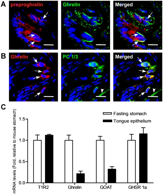
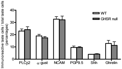
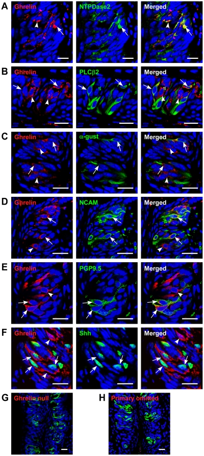
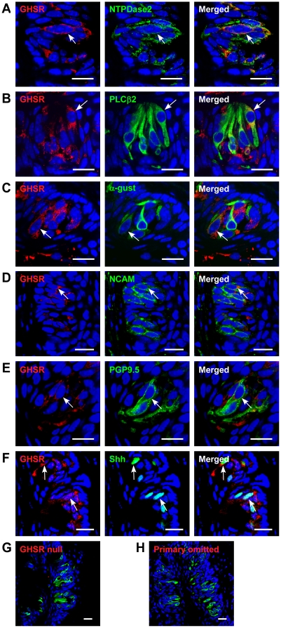
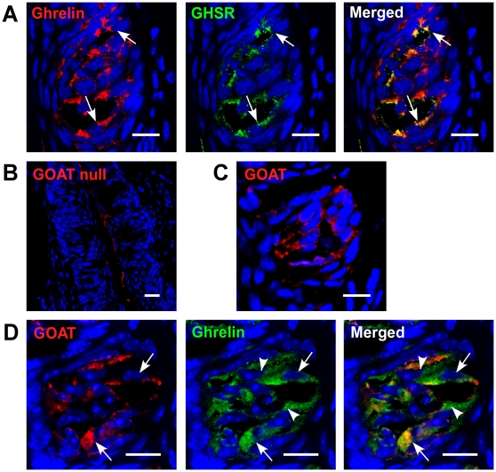
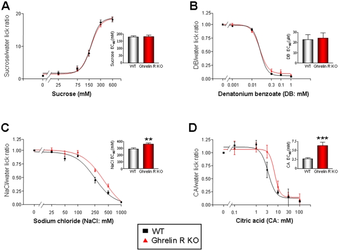
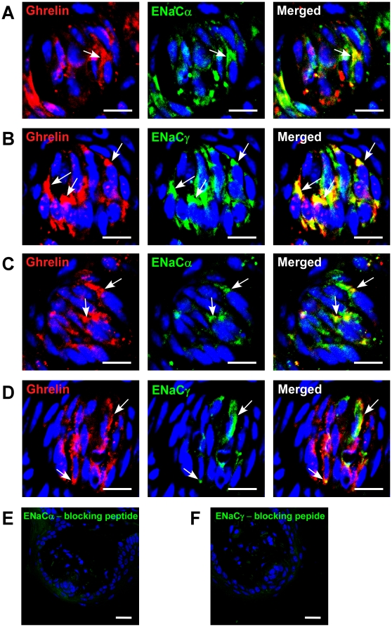
Methodology/principal findings: Using immunohistochemical analyses, we show that ghrelin, a hormone classically considered to be an appetite-regulating hormone, is present within the taste buds of the tongue. Prepro-ghrelin, prohormone convertase 1/3 (PC 1/3), ghrelin, its cognate receptor (GHSR), and ghrelin-O-acyltransferase (GOAT , the enzyme that activates ghrelin) are expressed in Type I, II, III and IV taste cells of mouse taste buds. In addition, ghrelin and GHSR co-localize in the same taste cells, suggesting that ghrelin works in an autocrine manner in taste cells. To determine a role for ghrelin in modifying taste perception, we performed taste behavioral tests using GHSR null mice. GHSR null mice exhibited significantly reduced taste responsivity to sour (citric acid) and salty (sodium chloride) tastants.

Conclusions/significance: These findings suggest that ghrelin plays a local modulatory role in determining taste bud signaling and function and could be a novel mechanism for the modulation of salty and sour taste responsivity.


Conflict of interest statement

Competing Interests: The authors have declared that no competing interests exist.

Figures

Figure 1
Figure 1. Expression of a prepro-ghrelin, PC1/3, and ghrelin in circumvallate papillae (CV) taste cells of mice.
(A) preproghrelin and ghrelin are co-localized in a subset of taste cells. Arrows, cells expressing both proteins. (B) ghrelin and PC1/3 are co-expressed in a subset of PC 1/3-positive cells. Arrowhead, cells expressing only PC 1/3; arrows, cells expressing both. Scale bars, 20 µm. Blue is TO-PRO-3 nuclear stain. (C) Quantitative real-time PCR of cRNA from tongue and stomach. Experiments were carried out in triplicate and replicated at least twice. Values are expressed as means ± S.E.M.
Figure 2
Figure 2. Comparison of cell types in circumvallate papillae of wild type and GHSR null mice.
The total number of cells in the section was determined by counting the number of TO-PRO-3 stained nuclei present in each taste bud. Percentage of immunoreactive taste cells was calculated by dividing the number of immunoreactive taste cells by the total number of the taste cells in each taste bud. Values are expressed as means ± S.E.M.
Figure 3
Figure 3. Co-expression of ghrelin and taste cell markers in circumvallate papillae (CV) of mice.
(A) ghrelin and NTPDase2 are co-localized in a subset of NTPDase2-positive cells. Arrowhead, cell expressing only ghrelin; arrow, cell expressing both. (B) ghrelin and PLCβ2 are co-localized in a subset of PLCβ2-positive cells. Arrowhead, cell expressing only ghrelin; arrow, cells expressing both. (C) ghrelin and α-gustducin are co-localized in a subset of α-gustducin-positive cells. Arrowhead, cell expressing only ghrelin; arrows, cell expressing both. (D) ghrelin and NCAM are co-localized in a subset of NCAM-positive cells. Arrowhead, cell expressing only ghrelin; arrow, cell expressing both. (E) ghrelin and PGP9.5 are co-localized in a subset of PGP9.5-positive cells. Arrowhead, cell expressing only ghrelin; arrow, cell expressing both. (F) ghrelin and Shh are co-localized in a subset of Shh-positive cells. Arrowhead, cell expressing only ghrelin; arrows, cells expressing both. (G) ghrelin and PLCβ2 are stained in ghrelin null mice as a negative control. (H) ghrelin and PLCβ2 are co-stained after the ghrelin antibody was omitted. Scale bars, 20 µm. Blue is TO-PRO-3 nuclear stain.
Figure 4
Figure 4. Co-expression of GHSR and taste cell markers in circumvallate papillae (CV) of mice.
(A) GHSR and NTPDase2 are co-localized in a subset of NTPDase2-positive cells (arrow). (B) GHSR and PLCβ2 are co-localized in a subset of PLCβ2-positive cells (arrow). (C) GHSR and α-gustducin are co-localized in a subset of α-gustducin-positive cells. Arrow, cells expressing both. (D) GHSR and NCAM are co-localized in a subset of NCAM-positive cells. Arrow, cell expressing both. (E) GHSR and PGP9.5 are co-localized in a subset of PGP9.5-positive cells (arrow). (F) GHSR and Shh are co-localized in a subset of Shh-positive cells (arrows). (G) GHSR and PLCβ2 are stained in ghrelin null mice as a negative control. (H) GHSR and PLCβ2 are co-stained after the ghrelin antibody was omitted. Scale bars, 20 µm. Blue is TO-PRO-3 nuclear stain.
Figure 5
Figure 5. Co-expression of ghrelin/GHSR and ghrelin/GOAT in circumvallate papillae (CV) of mice.
(A) Ghrelin and GHSR are co-localized in a subset of ghrelin-positive cells (arrows). (B) GOAT is stained in GOAT null mice as a negative control. (C) GOAT is expressed in TCs. (D) GOAT and ghrelin are co-localized in a subset of ghrelin-positive cells (arrows). Scale bars, 20 µm. Blue is TO-PRO-3 nuclear stain.
Figure 6
Figure 6. Altered salt and sour taste responses of WT and GHSR null mice in brief access taste tests.
Taste responses, expressed as tastant/water lick ratios and as a function of stimulus concentration, of GHSR null, (red) and WT (black) to (A) sucrose, (B) DB, (C) NaCl and (D) CA. Points are expressed as means ± S.E.M. Curves were fit as described in Methods. **p<0.01; ***p<0.001.
Figure 7
Figure 7. Co-expression of ghrelin and ENaC subunits in fungiform papillae of WT (A,B) and GHSR null (C,D) mice.
(A) ghrelin and ENaCα are co-localized in a subset of ghrelin-positive cells in WT mice. Arrow, cell expressing both. (B) ghrelin and ENaCγ are co-localized in a subset of ghrelin-positive cells in WT mice. Arrows, cells expressing both. (C) ghrelin and ENaCα are co-localized in a subset of ghrelin-positive cells in GHSR null mice. Arrows, cells expressing both. (D) ghrelin and ENaCγ are co-localized in a subset of ghrelin-positive cells in GHSR null mice. Arrows, cells expressing both. (E) ENaCα staining after treatment with blocking peptide. (F) ENaCγ staining after treatment with blocking peptide. Scale bars, 20 µm. Blue is TO-PRO-3 nuclear stain.

Similar articles

Cited by

References

    1. Scott K. Taste recognition: food for thought. Neuron. 2005;48:455–464. - PubMed
    1. Chandrashekar J, Hoon MA, Ryba NJ, Zuker CS. The receptors and cells for mammalian taste. Nature. 2006;444:288–294. - PubMed
    1. Roper SD. Cell communication in taste buds. Cell Mol Life Sci. 2006;63:1494–1500. - PMC - PubMed
    1. Pumplin DW, Yu C, Smith DV. Light and dark cells of rat vallate taste buds are morphologically distinct cell types. J Comp Neurol. 1997;378:389–410. - PubMed
    1. Tomchik SM, Berg S, Kim JW, Chaudhari N, Roper SD. Breadth of tuning and taste coding in mammalian taste buds. J Neurosci. 2007;27:10840–10848. - PMC - PubMed

Publication types