. 2010 Sep 14;5(9):e12723.
doi: 10.1371/journal.pone.0012723.

A new method for the characterization of strain-specific conformational stability of protease-sensitive and protease-resistant PrPSc

Affiliations

A new method for the characterization of strain-specific conformational stability of protease-sensitive and protease-resistant PrPSc

Laura Pirisinu et al. PLoS One. .

Abstract

Although proteinacious in nature, prions exist as strains with specific self-perpetuating biological properties. Prion strains are thought to be associated with different conformers of PrP(Sc), a disease-associated isoform of the host-encoded cellular protein (PrP(C)). Molecular strain typing approaches have been developed which rely on the characterization of protease-resistant PrP(Sc). However, PrP(Sc) is composed not only of protease-resistant but also of protease-sensitive isoforms. The aim of this work was to develop a protocol for the molecular characterization of both, protease-resistant and protease-sensitive PrP(Sc) aggregates. We first set up experimental conditions which allowed the most advantageous separation of PrP(C) and PrP(Sc) by means of differential centrifugation. The conformational solubility and stability assay (CSSA) was then developed by measuring PrP(Sc) solubility as a function of increased exposure to GdnHCl. Brain homogenates from voles infected with human and sheep prion isolates were analysed by CSSA and showed strain-specific conformational stabilities, with mean [GdnHCl](1/2) values ranging from 1.6 M for MM2 sCJD to 2.1 for scrapie and to 2.8 M for MM1/MV1 sCJD and E200K gCJD. Interestingly, the rank order of [GdnHCl](1/2) values observed in the human and sheep isolates used as inocula closely matched those found following transmission in voles, being MM1 sCJD the most resistant (3.3 M), followed by sheep scrapie (2.2 M) and by MM2 sCJD (1.6 M). In order to test the ability of CSSA to characterise protease-sensitive PrP(Sc), we analysed sheep isolates of Nor98 and compared them to classical scrapie isolates. In Nor98, insoluble PrP(Sc) aggregates were mainly protease-sensitive and showed a conformational stability much lower than in classical scrapie. Our results show that CSSA is able to reveal strain-specified PrP(Sc) conformational stabilities of protease-resistant and protease-sensitive PrP(Sc) and that it is a valuable tool for strain typing in natural hosts, such as humans and sheep.


Conflict of interest statement

Competing Interests: The authors have declared that no competing interests exist.

Figures

Figure 1
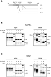
Figure 1. PrP species in healthy and scrapie-affected voles.
A: Schematic representation of full length PrP (FL) and PrP fragments (C1 and C2) generated by α and β cleavages. The location of SAF84, 12B2 and SAF32 epitopes used for the FL/C1/C2 discrimination are shown. B and C: Normal brain homogenates (NBH) and scrapie brain homogenates (SBH) of voles were analyzed by western blot using SAF84 (B) and 12B2 (C). The samples, were analyzed either before or after deglycosylation (N-Gly F) as indicated. The brackets on the left indicate the position of glycosylated and unglycosylated bands of FL, C2 and C1: from 35 kDa to 27 kDa for FL, from 26 kDa to 18 kDa for C2 and from 24 kDa to 16 kDa for C1. These PrP species are reduced to single unglycosylated PrP bands after deglycosylation, which are indicated by dashes on the right of the blots. In SBH, PrPSc dimers (Dim) are also indicated. In NBH both C1 and C2 were present, although C2 was poorly represented; in contrast in SBH C2 fragment was the most abundant PrP species while C1 was barely detectable. Tissue equivalents (TE) loaded per lane were 0,15 mg and 0.5 mg for samples respectively before and after PK digestion. Molecular weight markers were loaded into the last lane of each blot. The positions of MW markers are 15, 20, 25, 37 and 50 kDa.
Figure 2
Figure 2. Separation of PrPC and PrPSc in vole brain homogenates.
A: Western blot analysis of soluble and insoluble PrP fractions from normal brain homogenate (NBH) and scrapie brain homogenate (SBH) of voles. Samples were centrifuged at 20000 g for 1 h in presence of 2% sarcosyl, and supernatants (S) and pellets (P) were analysed before (+) and after (−) PK treatment. Aliquots of samples before centrifugation (Tot) were analysed too. TE per lane were 0,2 mg for “Tot” and “S” lanes, and 0.4 mg for “P” lanes. Brackets on the left indicate the position of the FL, C1 and C2 PrP fragments. Dimers (Dim) of PrPSc are indicated on the right in SBH blot. B: Western blot analysis of soluble and insoluble PrP fractions from scrapie brain homogenate (SBH) and an artificially mixed sample (SBH+NBH). Samples were centrifuged as described and total (Tot), supernatant (S) and pellet (P) fractions were deglycosylated. Full-length PrP (FL), C1 and C2 PrP fragments are indicated on the left. In each lane, 0.02 mg TE were loaded. C: Western blot analysis of soluble and insoluble PrP from brain homogenates of voles infected with MM1 and MM2 sCJD. The samples were treated as in panel A and total (Tot), supernatant (S) and pellet (P) fractions were analysed with or without PK treatment. In each lane 0.3 mg TE were loaded. Brackets on the left indicate the position of the FL, C1 and C2 fragments. Dimers (Dim) of PrPSc are indicated on the left. A–C: Membranes were probed with SAF84. Molecular size markers are shown in kilodaltons on the right of each panel.
Figure 3
Figure 3. Conformational stability and solubility assay in healthy and diseased voles.
A and B: Western blot analysis of scrapie brain homogenate (SBH) after denaturation with various concentrations of GdnHCl and separation of insoluble (A) and soluble (B) fractions by centrifugation. A: Immunoblot of pellets (P) at different concentrations of GdnHCl. In order to compare the fractions in the same blot, supernatant (S) at 0 M GdnHCl was loaded too. In the pellets, insoluble PrP decreased with increasing concentrations of GdnHCl (M). In each lane 0.4 mg TE were loaded. B: Immunoblot of supernatants at different concentrations of GdnHCl. In the first lane pellet (P) at 0 M GdnHCl was loaded too. In the supernatant PrP increased with increasing GdnHCl. In each lane 0.4 mg TE were loaded. Note that the FL and C1 PrP fragments are present in the supernatant after treatment with 0 M and 1 M GdnHCl, while the PrPSc-specific fragment C2 is visible in the supernatant from 1.5 M GdnHCl onwards, in parallel with the decrease of insoluble PrP in panel A. C: The conformational stability of PrPSc in SBH was analysed by denaturation curves best-fitted by plotting the fraction of PrPSc in the pellet (P) and in the supernatant (S), depicted in panel A and B respectively, as a function of GdnHCl concentration. [GdnHCl]1/2 values were 1.92 M in the pellet and 1.91 M in the supernatant fraction. D: Western blot analysis of supernatants (S) at different concentrations of GdnHCl from normal brain homogenate (NBH). In the first lane pellet (P) at 0 M GdnHCl was loaded. In NBH, PrPC was mostly in supernatant fraction and remained soluble at all GdnHCl concentrations tested. Samples were loaded as 0.4 mg tissue equivalent into each lane. A, B, D: Membranes were probed with SAF84. Molecular size markers are shown in kilodaltons on the left of each panel.
Figure 4
Figure 4. Relationship between PrPSc insolubility and resistance to PK.
The same SBH was analyzed in parallel by CSSA (A) and CSA (B), and the denaturation curves obtained were compared (C). A and B: Western blot analysis of insoluble PrPSc (A) and PK-resistant PrPSc (B) after denaturation of homogenates with increasing concentrations of GdnHCl. In each lane 0.2 mg TE were loaded. The concentrations of GdnHCl were: 0.5, 1.0, 1.5, 2.0, 2.5, 3.0 and 4.0 M. C: Graph depicting the denaturation curves obtained by CSSA (panel A) and CSA (panel B). [GdnHCl]1/2 values were 2.14 M and 2.23 M for CSSA and CSA, respectively. D: Western blot showing that PrPSc loses PK-resistance upon solubilization. In absence of treatment with GdnHCl (0), total PrP (Tot) from SBH was resistant to PK digestion (compare PK- and PK+ lanes). After treatment with 3 M GdnHCl (3) and differential centrifugation, most of PrPSc was found in the supernatant (S) and was also made PK-susceptible. In absence of treatment with GdnHCl (0), the supernatant (S) contained normal PrP which was susceptible to PK. Note the different banding patterns in the supernatants containing PrPC (0) and solubilised PrPSc (3). A, B, D: Membranes were probed with SAF84. Molecular size markers are shown in kilodaltons on the left of each panel.
Figure 5
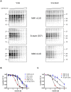
Figure 5. Conformational stability and solubility assay of different strains in voles and natural hosts.
A and B: Conformational stability of PrPSc from voles inoculated with MM1 sCJD, Scrapie and MM2 sCJD and from the respective human and sheep isolates. A: Representative western blots of insoluble PrPSc from voles (left column) and natural hosts (on the right column) after treatment of homogenates with increasing concentrations of GdnHCl. The concentrations of GdnHCl were: 0, 0.5, 1.0, 1.5, 2.0, 2.5, 3.0 and 4.0 M. TE loaded per lane were 0.2 mg for vole samples, 0.34 mg for human isolates and 0.125 mg for the scrapie isolate. Membranes were probed with SAF84 (vole samples and sheep scrapie) or with L42 (human isolates). B: Dose-response curves in vole strains, obtained by plotting the fraction of PrPSc remaining in the pellet as a function of GdnHCl concentration and best-fitted and using a four parameter logistic equation. Individual curves were combined within each strain group (Scrapie, E200K gCJD, MV1, MM1 and MM2 sCJD) in order to compare the denaturation profiles of the 5 groups. C: Dose-response curves from sheep and human isolates, obtained by plotting the fraction of PrPSc remaining in the pellet as a function of GdnHCl concentration and best-fitted and using a four parameter logistic equation. [GdnHCl]1/2 values of MM1, MM2 and scrapie isolates were 3,31 M, 1,63 M and 2,23 M respectively.
Figure 6
Figure 6. Separation of soluble and insoluble PrP fractions in classical scrapie and Nor98.
Western blot analysis of classical scrapie and Nor98 ovine isolates. Supernatant (S) and pellet (P) fractions were analysed with (+) or without (−) PK treatment. In each lane 0.8 mg TE were loaded. Molecular size markers are shown in kilodaltons on the left of the blot. Membrane was probed with L42.
Figure 7
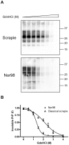
Figure 7. CSSA of classical scrapie and Nor98 isolates.
A: Representative western blots of insoluble PrP after denaturation with increasing concentrations of GdnHCl in classical scrapie (ARQ/AHQ) and Nor98 (ARR/AHQ) isolates. The concentrations of GdnHCl were: 0, 0.5, 1.0, 1.5, 2.0, 2.5, 3.0 and 4.0 M. TE loaded per lane were 0.125 mg for classical scrapie and 0.3 mg for Nor98. Membranes were probed with L42. Molecular size markers are shown in kilodaltons on the right of each blot. B: Dose-response curves of Nor98 and classical scrapie, obtained by plotting the fraction of PrPSc remaining in the pellet as a function of GdnHCl concentration and best-fitted and using a four parameter logistic equation. Individual curves were combined within each strain group (Scrapie and Nopr98) in order to compare the denaturation profiles of the two groups. Individual [GdnHCl]1/2 values are shown in Table 2.

Similar articles

Cited by

References

    1. Biacabe AG, Laplanche JL, Ryder S, Baron T. Distinct molecular phenotypes in bovine prion diseases. EMBO Rep. 2004;5:110–115. - PMC - PubMed
    1. Casalone C, Zanusso G, Acutis P, Ferrari S, Capucci L, et al. Identification of a second bovine amyloidotic spongiform encephalopathy: molecular similarities with sporadic Creutzfeldt-Jakob disease. Proc Natl Acad Sci U S A. 2004;101:3065–3070. - PMC - PubMed
    1. Benestad SL, Sarradin P, Thu B, Schonheit J, Tranulis MA, et al. Cases of scrapie with unusual features in Norway and designation of a new type, Nor98. Vet Rec. 2003;153:202–208. - PubMed
    1. Bolton DC, McKinley MP, Prusiner SB. Identification of a protein that purifies with the scrapie prion. Science. 1982;218:1309–1311. - PubMed
    1. Meyer RK, McKinley MP, Bowman KA, Braunfeld MB, Barry RA, et al. Separation and properties of cellular and scrapie prion proteins. Proc Natl Acad Sci U S A. 1986;83:2310–2314. - PMC - PubMed

Publication types