Clinical Trial
. 2010 Sep 14;5(9):e12728.
doi: 10.1371/journal.pone.0012728.

Colonic biopsies to assess the neuropathology of Parkinson's disease and its relationship with symptoms

Affiliations
Clinical Trial

Colonic biopsies to assess the neuropathology of Parkinson's disease and its relationship with symptoms

Thibaud Lebouvier et al. PLoS One. .

Abstract

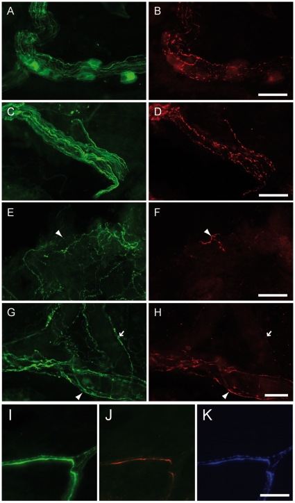
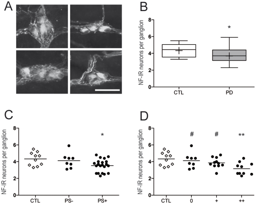
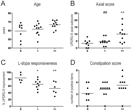
Background: The presence of Lewy bodies and Lewy neurites (LN) has been demonstrated in the enteric nervous system (ENS) of Parkinson's disease (PD) patients. The aims of the present research were to use routine colonoscopy biopsies (1) to analyze, in depth, enteric pathology throughout the colonic submucosal plexus (SMP), and (2) to correlate the pathological burden with neurological and gastrointestinal (GI) symptoms.

Methodology/principal findings: A total of 10 control and 29 PD patients divided into 3 groups according to disease duration were included. PD and GI symptoms were assessed using the Unified Parkinson's Disease Rating Scale part III and the Rome III questionnaire, respectively. Four biopsies were taken from the ascending and descending colon during the course of a total colonoscopy. Immunohistochemical analysis was performed using antibodies against phosphorylated alpha-synuclein, neurofilaments NF 220 kDa (NF) and tyrosine hydroxylase (TH). The density of LN, labeled by anti-phosphorylated alpha-synuclein antibodies, was evaluated using a quantitative rating score. Lewy pathology was apparent in the colonic biopsies from 21 patients and in none of the controls. A decreased number of NF-immunoreactive neurons per ganglion was observed in the SMP of PD patients compared to controls. The amount of LN in the ENS was inversely correlated with neuronal count and positively correlated with levodopa-unresponsive features and constipation.

Conclusion/significance: Analysis of the ENS by routine colonoscopy biopsies is a useful tool for pre-mortem neuropathological diagnosis of PD, and also provides insight into the progression of motor and non-motor symptoms.

Trial registration: ClinicalTrials.gov NCT00491062.


Conflict of interest statement

Competing Interests: The authors have declared that no competing interests exist.

Figures

Figure 1
Figure 1. Phospho-α-synuclein-positive submucosal neurites in PD patients.
Labeling with antibodies against neurofilament (NF) (ACEGI) and phosphorylated α-synuclein (BDFHJ) revealed that some NF-immunoreactive (IR) neurites were also phospho-α-synuclein-IR. Occasionally, these phospho-α-synuclein-IR neurites were present amidst a submucosal ganglion (AB). Some of these structures formed bundles (D) while others were isolated (arrowhead in F). Thirty seven percents of the phospho-α-synuclein-IR neurites were perivascular (GH). Triple immunostaining with antibodies against tyrosine hydroxylase (K) revealed that 60% of LN were also TH-immunoreactive (IJK). Scale bar: 30 µm.
Figure 2
Figure 2. Count of neurofilament-positive neurons in the submucosal plexus of PD patients.
A. Neurofilament-immunoreactive (NF-IR) submucosal neurons were counted in every available ganglion from colonic biopsies. Representative photographs of ganglia from PD patients (left panels) and controls (right panels). Scale bar: 30 µm. B. A significant decrease in the number of NF-IR neurons per ganglion was present in the SMP of PD patients (PD, n = 29) as compared to controls (CTL, n = 10) (p<0.05). The bottom and the top of the box represent the 25th and 75th percentiles, respectively, and the end of the whiskers represent the minimum and maximum values; the median is represented as a bar and the mean as a ‘+’ sign inside the box. C. When segregating patients according to the presence (PS+) or absence (PS-) of phospho-synuclein IR neurites, the difference between patients and controls was sustained only for the group with Lewy pathology (PS+, n = 21, p<0.05). D. When further stratifying patients according to the density of pathology, the difference between patients and controls was sustained only for the group with severe Lewy pathology (++, n = 10, p<0.01). Groups without (0) or with moderate pathology (+) significantly differed from the group with severe (++) pathology (p<0.05). Each white square represents one control, each black circle represents one PD patient. Horizontal bars represent the mean. *p<0.05 and **p<0.01 as compared with controls. #p<0.05 as compared with the group with severe pathology (++).
Figure 3
Figure 3. Correlation of clinical symptoms with pathology burden.
A. Measure of pathology burden using a quantitative score correlated with age, which appeared as a potential confounding factor. Subsequent correlation analysis was performed after adjusting the data for age. B. Pathology burden positively correlated with axial score, which measures axial symptoms such as dysarthria and postural instability. The group with severe pathology (++) significantly differed from the group with absent (0) or moderate (+) pathology (p<0.01). C. Pathology burden also correlated with L-Dopa responsiveness, estimated by the percentage of UPDRS-III improvement after L-Dopa intake. Responsiveness was higher in the group with absent pathology (0), as compared with the group with moderate (+, p<0.05) or severe (++, p<0.01) pathology. D. Pathology burden also correlated with constipation severity, as defined by the number of positive answers to the constipation items of Rome III questionnaire. Each black circle represents one PD patient. Horizontal bars represent the mean. *p<0.05 and **p<0.01 as compared with the group with absent pathology (0). #p<0.05 and ##p<0.01 as compared with the group with severe pathology (++).

References

    1. Furness JB. The enteric nervous system: normal functions and enteric neuropathies. Neurogastroenterol Motil. 2008;20(Suppl 1):32–38. - PubMed
    1. De Giorgio R, Camilleri M. Human enteric neuropathies: morphology and molecular pathology. Neurogastroenterol Motil. 2004;16:515–531. - PubMed
    1. Basilisco G, Gebbia C, Peracchi M, Velio P, Conte D, et al. Cerebellar degeneration and hearing loss in a patient with idiopathic myenteric ganglionitis. Eur J Gastroenterol Hepatol. 2005;17:449–452. - PubMed
    1. Haik S, Faucheux BA, Hauw JJ. Brain targeting through the autonomous nervous system: lessons from prion diseases. Trends Mol Med. 2004;10:107–112. - PubMed
    1. Joiner S, Linehan JM, Brandner S, Wadsworth JD, Collinge J. High levels of disease related prion protein in the ileum in variant Creutzfeldt-Jakob disease. Gut. 2005;54:1506–1508. - PMC - PubMed

Publication types

Associated data