. 2010 Sep 14;5(9):e12743.
doi: 10.1371/journal.pone.0012743.

Gingival fibroblasts as a promising source of induced pluripotent stem cells

Affiliations

Gingival fibroblasts as a promising source of induced pluripotent stem cells

Hiroshi Egusa et al. PLoS One. .

Abstract

Background: Induced pluripotent stem (iPS) cells efficiently generated from accessible tissues have the potential for clinical applications. Oral gingiva, which is often resected during general dental treatments and treated as biomedical waste, is an easily obtainable tissue, and cells can be isolated from patients with minimal discomfort.

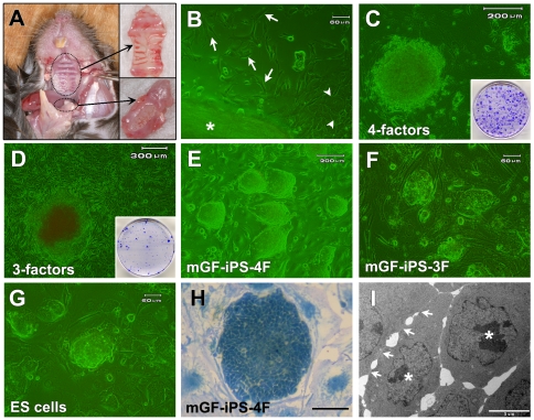
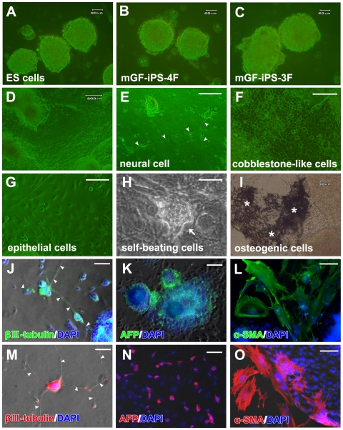
Methodology/principal findings: We herein demonstrate iPS cell generation from adult wild-type mouse gingival fibroblasts (GFs) via introduction of four factors (Oct3/4, Sox2, Klf4 and c-Myc; GF-iPS-4F cells) or three factors (the same as GF-iPS-4F cells, but without the c-Myc oncogene; GF-iPS-3F cells) without drug selection. iPS cells were also generated from primary human gingival fibroblasts via four-factor transduction. These cells exhibited the morphology and growth properties of embryonic stem (ES) cells and expressed ES cell marker genes, with a decreased CpG methylation ratio in promoter regions of Nanog and Oct3/4. Additionally, teratoma formation assays showed ES cell-like derivation of cells and tissues representative of all three germ layers. In comparison to mouse GF-iPS-4F cells, GF-iPS-3F cells showed consistently more ES cell-like characteristics in terms of DNA methylation status and gene expression, although the reprogramming process was substantially delayed and the overall efficiency was also reduced. When transplanted into blastocysts, GF-iPS-3F cells gave rise to chimeras and contributed to the development of the germline. Notably, the four-factor reprogramming efficiency of mouse GFs was more than 7-fold higher than that of fibroblasts from tail-tips, possibly because of their high proliferative capacity.

Conclusions/significance: These results suggest that GFs from the easily obtainable gingival tissues can be readily reprogrammed into iPS cells, thus making them a promising cell source for investigating the basis of cellular reprogramming and pluripotency for future clinical applications. In addition, high-quality iPS cells were generated from mouse GFs without Myc transduction or a specific system for reprogrammed cell selection.


Conflict of interest statement

Competing Interests: The authors have declared that no competing interests exist.

Figures

Figure 1
Figure 1. Generation of mouse GF-derived iPS cells.
(A) Palatal (upper inset) and mandibular (lower inset) gingival tissues of adult mice were extracted for establishment of primary GFs. (B) Fibroblasts (arrow) and epithelial cells (arrowhead) migrated out of the palatal gingival tissue (asterisk). Scale bar; 60 µm. (C) The morphology of a colony derived from mGFs 19 days after transduction of the four factors. Scale bar; 200 µm. Inset: Colonies in a 60-mm dish after staining with crystal violet (CV) on day 21. (D) The morphology of a colony derived from mGFs 49 days after transduction of the three factors. Scale bar; 300 µm. Inset: Colonies in a 100-mm dish after staining with CV on day 50. (E–G) Morphology of (E) mGF-iPS-4F-1 cells (Scale bar; 200 µm), (F) mGF-iPS-3F-1 cells (Scale bar; 60 µm) and (G) mouse ES cell line (Scale bar; 60 µm). (H) Morphology of a pGF-iPS-4F-1 colony stained with methylene blue. Scale bar; 50 µm (I) TEM photograph of a pGF-iPS-4F-1 cell colony showing tight and continual cell membrane contacts (arrows), large nucleoi (asterisks) and scant cytoplasm. Scale bar; 5 µm.
Figure 2
Figure 2. Characteristics of the mouse GF-iPS cells.
(A) GF-iPS cell colonies (upper panels: pGF-iPS-4F-1, lower panels: mGF-iPS-3F-4) positively stained for ALP. Scale bar; 30 µm. (B) Bisulfite sequencing of the Nanog and Oct3/4 promoters revealed that CpGs in the parental mGFs were converted to a demethylated state in the GF-iPS cells induced by the three or four factors, resulting in a methylation pattern similar to that of mouse ES cells. The numbers in the panel indicate CpG loci respective to the transcription start site (TSS) of the genes (blue: untranslated region, yellow: translated region). (C and D) RT-PCR analysis of ES cell marker genes (Nanog, ERas, Rex1) and endogenous Oct3/4, Sox2, Klf4 and c-Myc genes in GF-iPS-4F clones (C) or GF-iPS-3F clones (D), mouse ES cells, parental mGFs and SNL feeder cells. GAPDH was used as a loading control.
Figure 3
Figure 3. In vitro differentiation of mouse GF-iPS cells.
(A–C) In vitro EB formation of (A) mouse ES cells, (B) pGF-iPS-4F-2 cells and (C) mGF-iPS-3F-9 cells after 3 days in floating culture. Scale bars; 60 µm. (D) Morphology of pGF-iPS-4F-3 cells cultured on gelatin-coated plates without feeder cells for 10 days. Scale bar; 200 µm. Attached cells showed various morphologies, such as those resembling (E) neural cells (arrowheads: neurite-like outgrowth), (F) cobblestone-like cells and (G) epithelial cells. Scale bars; 100 µm. (H) Beating myocardial cells (arrow) in the pGF-iPS-4F-3 culture after 20 days of expansion. Scale bar; 100 µm. (I) Osteogenic cells with mineralized nodule formation (asterisks) in the pGF-iPS-4F-1 cell culture detected by von Kossa staining after 30 days of expansion. Scale bar; 30 µm. (J–L) pGF-iPS-4F-1 cells and (M–O) mGF-iPS-3F-2 cells were specifically directed to differentiate into cells from all three germ layers at days three and ten after expansion, respectively. (J and M) β-III tubulin-positive ectodermal neural cells (arrowheads: neurite-like outgrowth). (K and N) AFP-positive endodermal hepatic cells. (L and O) α-SMA-positive mesodermal smooth muscle cells. Nuclei are stained with DAPI. Scale bars; 200 µm.
Figure 4
Figure 4. Teratoma formation and germline chimeras from mouse GF-iPS cells.
(A) Transplantation of mGF-iPS-3F-1 and pGF-iPS-4F-1 cells into mouse testes resulted in apparent teratoma formation (dotted circle: tumor formation by pGF-iPS-4F-1 cell transplantation at week 10). Insets: Extracted teratomas from mice transplanted with pGF-iPS-4F-1 (upper) or mGF-iPS-3F-1 (lower) cells. Scale bars; 1 cm. (B–G) H&E staining of teratoma sections showed differentiation of mGF-iPS-3F-1 cells into various tissues from all three germ layers, including keratin-containing epidermal tissues (B: ectoderm), neural tissues (C: ectoderm), striated muscle (D: mesoderm), cartilage (E: mesoderm), adipose tissues (F: mesoderm) and gut-like epithelial tissues (G: endoderm). Scale bars; 100 µm. (H) Chimeric mice generated by injecting the black mouse-derived mGF-iPS-3F-1 cells into white mouse-derived blastocyst embryos. (I) An adult old chimeric male mouse generated from mGF-iPS-3F-3 cells (arrow head) was mated with Jcl:MCH white female mice and achieved germline transmission, as indicated by coat color in black (arrow). Inset: A newborn mouse generated via germline transmission (arrow).
Figure 5
Figure 5. Reprogramming efficiency of mouse GF- and TTF-derived iPS cells.
(A) Fibroblasts established from palatal (pGFs) and mandibular (mGFs) gingival tissues as well as tail-tips (TTFs) of the same mouse were simultaneously induced into iPS cells by four-factor transduction. Left panels: ES cell-like colony formation from each cell type at passage 4 was determined by ALP staining (Scale bars; 200 µm). Right panel: The reprogramming efficiency at passages 4, 7 and 10 was calculated as the number of ALP-stained iPS colonies formed per number of infected cells seeded. The data represent the mean values ± s.d. (n = 4). Significant differences (*P<0.01: ANOVA with Dunnett's correction for multiple comparisons) were evaluated with respect to the values for TTF at each passage number of cultures. (B) A cell proliferation assay was performed on the pGF, mGF and TTF cultures at passage 5. The data represent the mean values ± s.d. (n = 3). Significant differences (*P<0.01: ANOVA with Dunnett's correction for multiple comparisons) were evaluated with respect to the values for TTF at each time point. (C) Real-time RT-PCR analysis for endogenous expression of c-Myc, Klf4, Sox2, p53, p21 and Tert genes in pGFs, mGFs and TTFs at passages 4 and 6 (left panel). The expression level of Tert mRNA in pGFs was maintained for 6 passages, while that in TTFs decreased as the passage number increased (right panel). Expression of GAPDH was used as an internal control. The data represent the relative mRNA expression levels with respect to the expression levels of each gene in TTFs at passage 4.
Figure 6
Figure 6. Generation of human GF-derived iPS cells.
(A) Resection of gingival tissue from the adult patient during dental implant surgery. The resected gingival tissue (inset: scale bar; 1 mm) is generally treated as biomedical waste. (B) Fibroblasts (arrow) and epithelial cells (arrowhead) migrated out of the human gingival tissue (asterisk). Scale bar; 100 µm. (C) The morphology of established hGFs (Passage 4). Scale bar; 60 µm. (D) The morphology of a colony derived from hGFs 26 days after transduction of the four factors. Scale bar; 500 µm. (E–I) Morphology of (E and F) clonal hGF-derived iPS cells (clone 547A-1), (G) human dermal fibroblast-derived iPS cells (DF-iPS cells) and (H and I) human KhES-3 ES cells. Scale bars; 500 µm for E, G and H, and 50 µm for F and I.
Figure 7
Figure 7. Characteristics and differentiation of the hGF-iPS cells.
(A) hGF-iPS cell colonies positively stained for ALP (clone 547A-1). Scale bar; 300 µm. (B) RT-PCR analysis of ES cell specific genes (NANOG, REX1, TERT, endogenous OCT3/4 and SOX2) in hGF-iPS clones, human ES cell lines (H9 and KhES-3), parental hGFs and SNL feeder cells. GAPDH was used as a loading control. (C) Bisulfite sequencing of the NANOG and OCT3/4 promoters demonstrated that CpGs in the parental hGFs were converted to a demethylated state in the hGF-iPS cells (clones 547A-4 and -5), resulting in a methylation pattern similar to that of human H9 ES cells. (D–F) H&E staining of teratoma sections showed differentiation of hGF-iPS-547A-3 cells into neural tissues (D: ectoderm), cartilage (E: mesoderm), and gut-like epithelial tissues (F: endoderm). Scale bars; 50 µm.

Similar articles

Cited by

References

    1. Takahashi K, Yamanaka S. Induction of pluripotent stem cells from mouse embryonic and adult fibroblast cultures by defined factors. Cell. 2006;126:663–676. - PubMed
    1. Liu H, Zhu F, Yong J, Zhang P, Hou P, et al. Generation of induced pluripotent stem cells from adult rhesus monkey fibroblasts. Cell Stem Cell. 2008;3:587–590. - PubMed
    1. Shimada H, Nakada A, Hashimoto Y, Shigeno K, Shionoya Y, et al. Generation of canine induced pluripotent stem cells by retroviral transduction and chemical inhibitors. Mol Reprod Dev. 2010;77:2. - PubMed
    1. Ezashi T, Telugu BP, Alexenko AP, Sachdev S, Sinha S, et al. Derivation of induced pluripotent stem cells from pig somatic cells. Proc Natl Acad Sci U S A. 2009;106:10993–10998. - PMC - PubMed
    1. Takahashi K, Tanabe K, Ohnuki M, Narita M, Ichisaka T, et al. Induction of pluripotent stem cells from adult human fibroblasts by defined factors. Cell. 2007;131:861–872. - PubMed

Publication types