. 2010 Sep 14;5(9):e12735.
doi: 10.1371/journal.pone.0012735.

Prohibitins are required for cancer cell proliferation and adhesion

Affiliations

Prohibitins are required for cancer cell proliferation and adhesion

Claudia Sievers et al. PLoS One. .

Abstract

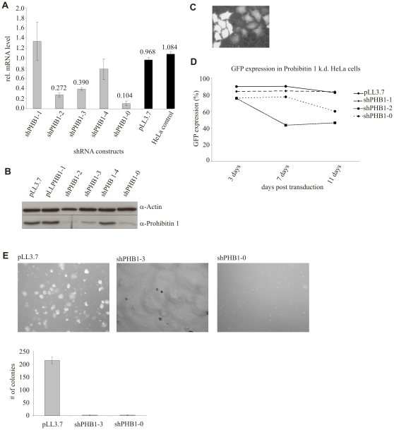
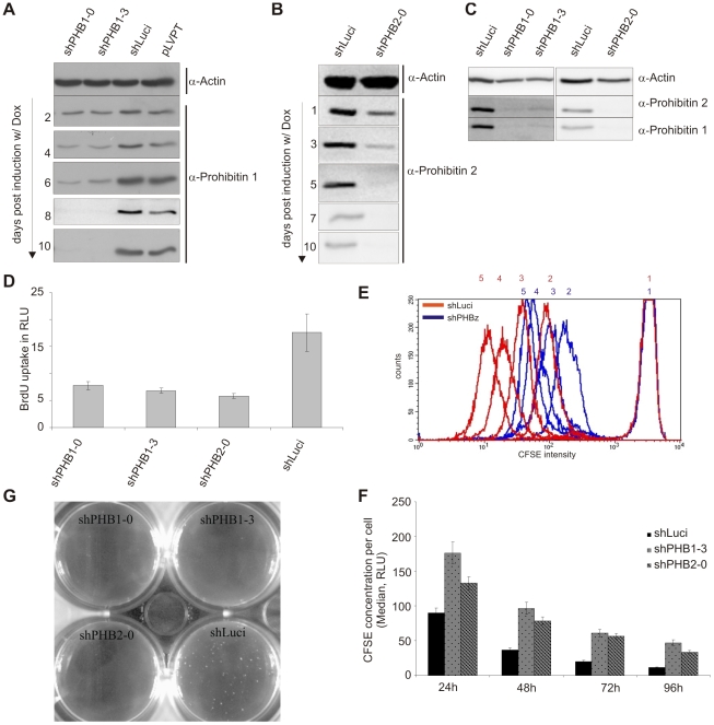
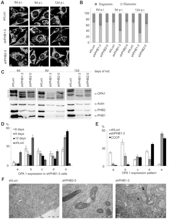
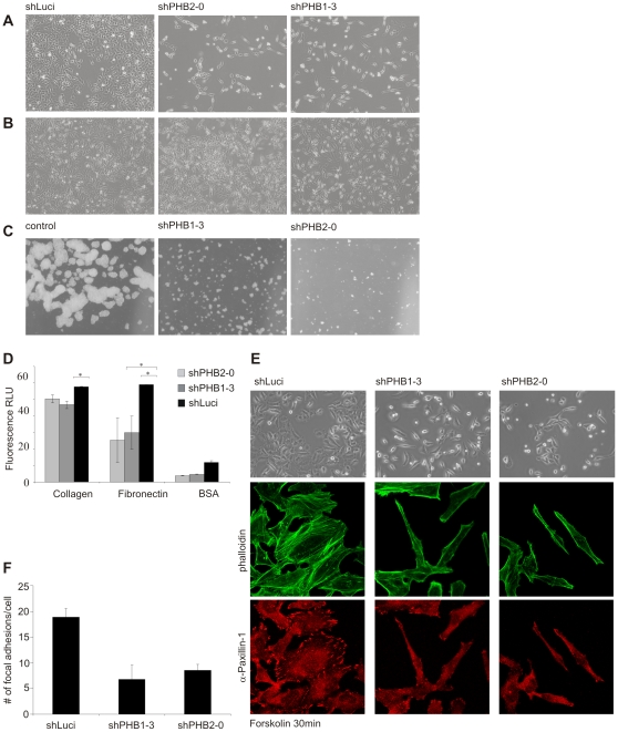
Prohibitin 1 (PHB1) is a highly conserved protein that together with its homologue prohibitin 2 (PHB2) mainly localizes to the inner mitochondrial membrane. Although it was originally identified by its ability to inhibit G1/S progression in human fibroblasts, its role as tumor suppressor is debated. To determine the function of prohibitins in maintaining cell homeostasis, we generated cancer cell lines expressing prohibitin-directed shRNAs. We show that prohibitin proteins are necessary for the proliferation of cancer cells. Down-regulation of prohibitin expression drastically reduced the rate of cell division. Furthermore, mitochondrial morphology was not affected, but loss of prohibitins did lead to the degradation of the fusion protein OPA1 and, in certain cancer cell lines, to a reduced capability to exhibit anchorage-independent growth. These cancer cells also exhibited reduced adhesion to the extracellular matrix. Taken together, these observations suggest prohibitins play a crucial role in adhesion processes in the cell and thereby sustaining cancer cell propagation and survival.


Conflict of interest statement

Competing Interests: The authors have declared that no competing interests exist.

Figures

Figure 1
Figure 1. Silencing of PHB1 led to a decrease in cell proliferation and anchorage-dependent growth in HeLa cells.
(A, B) PHB1 mRNA and protein levels were efficiently reduced in cells expressing shPHB1-0, shPHB1-2 and shPHB1-3 using the constitutive lentiviral expression system pLL3.7 as was shown by qRT-PCR and Western Blots, respectively. (C) Upon lentiviral transduction stable integration is indicated by GFP expression. (D) With the expression of positively validated shRNAs targeting PHB1 mRNA the fraction of GFP expressing cells was reduced within ten days of transduction from a pool of transduced cells within, as shown by FACS analysis. (E) Transduced HeLa cells expressing control vector or PHB1 targeting shRNAs were seeded in soft agar. Only control cells grew under anchorage-independent conditions and formed colonies.
Figure 2
Figure 2. Inducible expression of shRNAs targeting either PHB1 or PHB2 led to depletion of both proteins and subsequently to a slower cell proliferation rate.
(A, B) Transduction with the pLVPT lentiviral system allows doxycycline inducible shRNA expression. Within the indicated time of induction PHB1 and PHB2 protein expression was efficiently reduced. (C) The inducible expression of either PHB1 targeted or PHB2 targeted mRNA led to a reduced expression of both proteins, as shown by Western blot. (D) BrdU incorporation analysis shows HeLa cells expressing shPHB1-0, shPHB1-3 or shPHB2-0 for 10 days exhibit a reduced proliferation rate in comparison to cells expressing control shRNA (shLuci). (E) CFSE intensity as measured by FACS at 0 h (1), 24 h (2), 48 h (3), 72 h (4), and 96 h (5) is higher in prohibitin knockdown cells indicating reduced dilution of CFSE to fast replicating daughter cells. (F) CFSE intensity was gauged by FACS every 24 h for 5 days and the median of each peak was plotted against each other. (G) Inducible expression of shRNAs resulted in the same phenotype of reduced/loss of anchorage-independent growth in PHB1 (shPHB1-0, shPHB1-3) and PHB2 (shPHB2-0) knockdown cells as observed with the constitutive expression system.
Figure 3
Figure 3. Prohibitin knockdown led to fragmentation of the mitochondrial network without affecting cristae morphology.
(A, B) Mitochondria from prohibitin knockdown cells were stained with anti-Tom 20 and mitochondrial fragmentation was quantified using ImageJ software. An extended induction with doxycycline led to an increased fragmentation in shPHB1-3 expressing cells. (C) Western blots showing that prolonged doxycycline-induced shPHB1-3 expression led to a stronger prohibitin knockdown than shPHB2-0 expression, correlating with an increasing fragmentation of the fusion competent fragments a and b of OPA1. (D) Quantification showed that the fusion competent forms were degraded into the smaller fragments c and e. (E) Quantification of OPA1 patterning in control cells (shLuci), PHB1-depleted cells (shPHB1-3) and cells treated with 60 µM CCCP for 80 min (CCCP) to induce loss of ΔΨm. The OPA1 pattern in CCCP-treated cells differed from shPHB1-3 expressing cells, as the fragments a and b were completely lost while the smaller fragments d and e were increased. (F) TEM pictures of cells with a prohibitin knockdown induced for 10 days showed a more dense appearance of mitochondria than shLuci-expressing control cells, but the morphology of the cristae structure was not changed.
Figure 4
Figure 4. Prohibitin knockdown led to a changed morphology in HeLa cells and to reduced cell-cell contact.
(A, B) Silencing prohibitin expression for 10 days resulted in a stretched cell morphology with the nucleus slightly raised. (A) When seeded at low density prohibitin knockdown cells remained as single cells with minimal cell-cell contact. (B) At higher seed density, prohibitin-knockdown cells still showed an elongated morphology but either formed clusters (shPHB1-3) or remained predominantly as single cells with reduced cell-cell contact. The proliferation rate of prohibitin knockdown cells was also increased in comparison to cells seeded at low to normal density. (C) Preventing anchorage by seeding cells on agar-coated tissue culture plates strongly reduced proliferation rate and colony formation in prohibitin knockdown cells, whereas control cells (shLuci expressing cells) formed large cell clumps. (D) Prohibitin-depleted and control cells were treated with CFSE to increase fluorescence intensity and seeded on extracellular matrix proteins collagen or fibronectin, or on BSA for 30 min. Non- adherent cells were washed off and fluorescence intensity was measured. Prohibitin knockdown significantly reduced adhesion. (E) Doxycycline-induced cells were seeded on coverslips for 20 h followed by serum starvation for 4 h. After treatment with 50 µM Forskolin for 20 min, brightfield images showed lamellipodia formation in control cells but not in prohibitin- depleted cells. Furthermore, prohibitin-depleted cells showed decreased formation of focal adhesions as shown by immunocytochemistry staining using phalloidin and anti-paxillin antibodies. (F) For the quantification of focal adhesions in control and prohibitin knockdown cells, α-paxillin 1 stained spots were counted in 30 cells per condition.

Similar articles

Cited by

References

    1. McClung JK, Jupe ER, Liu XT, Dell'Orco RT. Prohibitin: potential role in senescence, development, and tumor suppression. Exp Gerontol. 1995;30:99–124. - PubMed
    1. Mishra S, Murphy LC, Murphy LJ. The Prohibitins: emerging roles in diverse functions. J Cell Mol Med. 2006;10:353–363. - PMC - PubMed
    1. Nijtmans LG, Artal SM, Grivell LA, Coates PJ. The mitochondrial PHB complex: roles in mitochondrial respiratory complex assembly, ageing and degenerative disease. Cell Mol Life Sci. 2002;59:143–155. - PMC - PubMed
    1. Terashima M, Kim KM, Adachi T, Nielsen PJ, Reth M, et al. The IgM antigen receptor of B lymphocytes is associated with prohibitin and a prohibitin-related protein. EMBO J. 1994;13:3782–3792. - PMC - PubMed
    1. Wang S, Fusaro G, Padmanabhan J, Chellappan SP. Prohibitin co-localizes with Rb in the nucleus and recruits N-CoR and HDAC1 for transcriptional repression. Oncogene. 2002;21:8388–8396. - PubMed

Publication types