

Myosin Va participates in acrosomal formation and nuclear morphogenesis during spermatogenesis of Chinese mitten crab Eriocheir sinensis
- PMID: 20856877
- PMCID: PMC2939076
- DOI: 10.1371/journal.pone.0012738
Myosin Va participates in acrosomal formation and nuclear morphogenesis during spermatogenesis of Chinese mitten crab Eriocheir sinensis
Abstract
Background: The Chinese mitten crab Eriocheir sinensis belongs to the Class Crustacea, Decapoda, Brachyura. The spermatozoon of this species is of aflagellated type, it has a spherical acrosome surrounded by the cup-shaped nucleus, which are unique to brachyurans. For the past several decades, studies on the spermatogenesis of the mitten crab mainly focus on the morphology. Compared with the extensive study of molecular mechanism of spermatogenesis in mammals, relatively less information is available in crustacean species. Myosin Va, a member of Class V myosin, has been implicated in acrosome biogenesis and vesicle transport during spermatogenesis in mammals. In the present study we demonstrate the expression and cellular localization of myosin Va during spermatogenesis in E. sinensis.
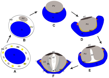
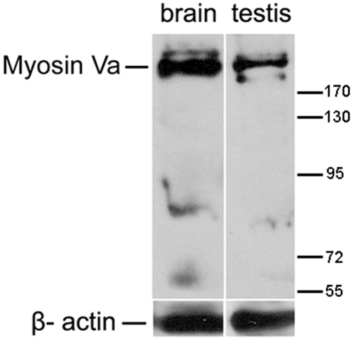
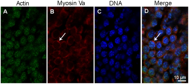
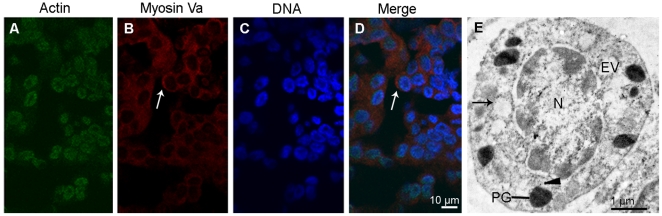
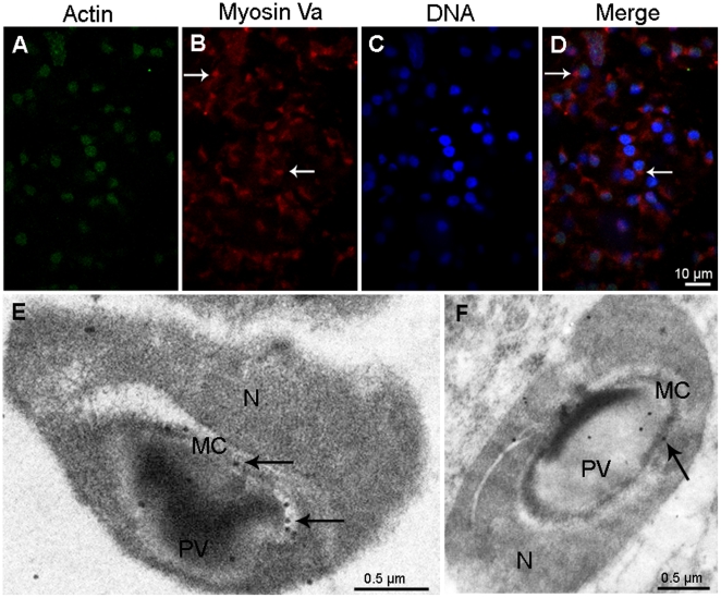
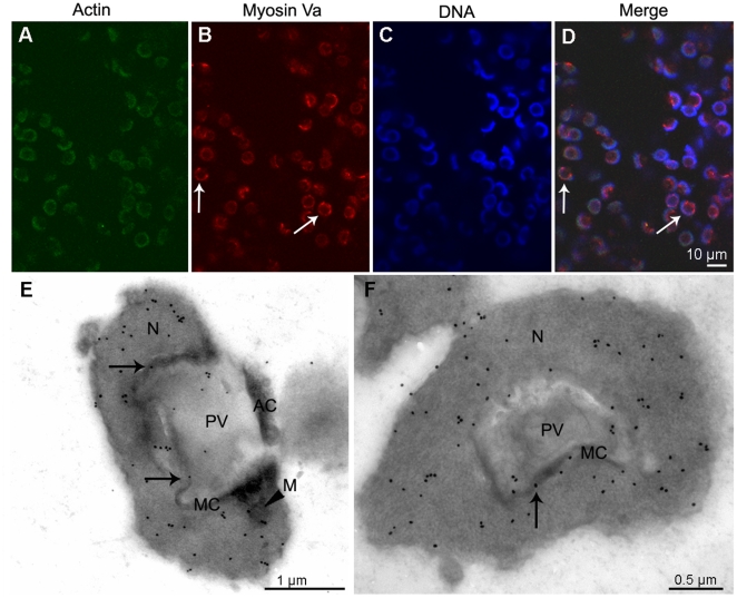
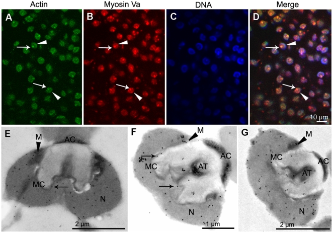
Methodology/principal findings: Western blot demonstrated that myosin Va is expressed during spermatogenesis. Immunocytochemical and ultrastructural analyses showed that myosin Va mainly localizes in the cytoplasm in spermatocytes. At the early stage of spermiogenesis, myosin Va binds to the endoplasmic reticulum vesicle (EV) and proacrosomal granule (PG). Subsequently, myosin Va localizes within the proacrosomal vesicle (PV) formed by PG and EV fusion and locates in the membrane complex (MC) at the mid spermatid stage. At the late spermatid stage, myosin Va is associated with the shaping nucleus and mitochondria. In mature spermatozoon, myosin Va predominates in acrosomal tubule (AT) and nucleus.
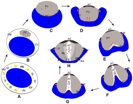
Conclusions/significance: Our study demonstrates that myosin Va may be involved in acrosome biogenesis and nuclear morphogenesis during spermatogenesis in E. sinensis. Considering the distribution and molecular characteristics of myosin Va, we also propose a hypothesis of AT formation in this species. It is the first time to uncover the role of myosin Va in crustacean spermatogenesis.
Conflict of interest statement
Figures
Similar articles
-
KIFC1 participates in acrosomal biogenesis, with discussion of its importance for the perforatorium in the Chinese mitten crab Eriocheir sinensis.Cell Tissue Res. 2009 Jul;337(1):113-23. doi: 10.1007/s00441-009-0800-3. Epub 2009 May 12. Cell Tissue Res. 2009. PMID: 19484267
-
Gene expression pattern of myosin Va during spermatogenesis of Chinese mitten crab, Eriocheir sinensis.Gene. 2012 Oct 15;508(1):78-84. doi: 10.1016/j.gene.2012.07.035. Epub 2012 Jul 27. Gene. 2012. PMID: 22846366
-
KIFC1 and myosin Va: two motors for acrosomal biogenesis and nuclear shaping during spermiogenesis of Portunus trituberculatus.Cell Tissue Res. 2017 Sep;369(3):625-640. doi: 10.1007/s00441-017-2638-4. Epub 2017 Jun 21. Cell Tissue Res. 2017. PMID: 28639134
-
Myosin superfamily: The multi-functional and irreplaceable factors in spermatogenesis and testicular tumors.Gene. 2016 Jan 15;576(1 Pt 2):195-207. doi: 10.1016/j.gene.2015.10.022. Epub 2015 Oct 19. Gene. 2016. PMID: 26478466 Review.
-
The acrosome-acroplaxome-manchette complex and the shaping of the spermatid head.Arch Histol Cytol. 2004 Nov;67(4):271-84. doi: 10.1679/aohc.67.271. Arch Histol Cytol. 2004. PMID: 15700535 Review.
Cited by
-
Transcriptome using Illumina sequencing reveals the traits of spermatogenesis and developing testes in Eriocheir sinensis.PLoS One. 2017 Feb 17;12(2):e0172478. doi: 10.1371/journal.pone.0172478. eCollection 2017. PLoS One. 2017. PMID: 28212420 Free PMC article.
-
ERK is involved in the process of acrosome reaction in vitro of the Chinese mitten crab, Eriocheir sinensis.Mar Biotechnol (NY). 2015 Jun;17(3):305-16. doi: 10.1007/s10126-015-9619-y. Epub 2015 Feb 8. Mar Biotechnol (NY). 2015. PMID: 25663286
-
Transcriptome Sequencing Reveals the Traits of Spermatogenesis and Testicular Development in Large Yellow Croaker (Larimichthys crocea).Genes (Basel). 2019 Nov 21;10(12):958. doi: 10.3390/genes10120958. Genes (Basel). 2019. PMID: 31766567 Free PMC article.
-
Molecular characterization and expression analysis of a KIFC1-like kinesin gene in the testis of Eumeces chinensis.Mol Biol Rep. 2013 Sep 29. doi: 10.1007/s11033-013-2779-9. Online ahead of print. Mol Biol Rep. 2013. PMID: 24078165
-
Myosin Va plays essential roles in maintaining normal mitosis, enhancing tumor cell motility and viability.Oncotarget. 2017 May 17;8(33):54654-54671. doi: 10.18632/oncotarget.17920. eCollection 2017 Aug 15. Oncotarget. 2017. PMID: 28903372 Free PMC article.
References
-
- Clermont Y. Kinetics of spermatogenesis in mammals: seminiferous epithelium cycle and spermatogonial renewal. Physiol Rev. 1972;52:198–236. - PubMed
-
- Pudney J. Spermatogenesis in nonmammalian vertebrates. Microsc Res Tech. 1995;32:459–497. - PubMed
-
- Du NS, Xue LZ, Lai W. Studies on the sperm of Chinese mitten-handed crab, Eriocheir sinensis (Crustacea, Decapoda). II. Spermatogenesis. Oceanol Limnol Sin. 1988;19:71–75.
-
- Yang WX, Du NS, Lai W. Changes of Golgi apparatus during spermatogenesis of Macrobrachium nipponense. Acta Zoologica Sinica. 1998;44:377–383.
-
- Yang WX, Du NS, Lai W. Junctional relationship between spermatogenic cell and Sertoli cells of freshwater shrimp, Macrobrachium nipponense. Acta Zoologica Sinica. 1999;45:178–186.
Publication types
MeSH terms
Substances
LinkOut - more resources
Full Text Sources
Miscellaneous
