

PET imaging of αvβ₃ integrin expression in tumours with ⁶⁸Ga-labelled mono-, di- and tetrameric RGD peptides
- PMID: 20857099
- PMCID: PMC3005123
- DOI: 10.1007/s00259-010-1615-x
PET imaging of αvβ₃ integrin expression in tumours with ⁶⁸Ga-labelled mono-, di- and tetrameric RGD peptides
Abstract
Purpose: Due to the restricted expression of α(v)β(3) in tumours, α(v)β(3) is considered a suitable receptor for tumour targeting. In this study the α(v)β(3)-binding characteristics of (68)Ga-labelled monomeric, dimeric and tetrameric RGD peptides were determined and compared with their (111)In-labelled counterparts.
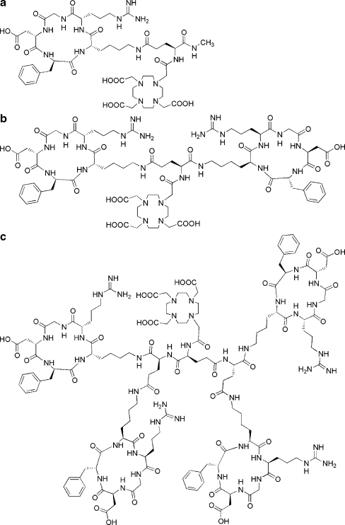
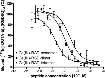
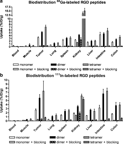
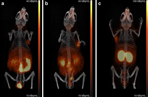
Methods: A monomeric (E-c(RGDfK)), a dimeric (E-[c(RGDfK)](2)) and a tetrameric (E{E[c(RGDfK)](2)}(2)) RGD peptide were synthesised, conjugated with DOTA and radiolabelled with (68)Ga. In vitro α(v)β(3)-binding characteristics were determined in a competitive binding assay. In vivo α(v)β(3)-targeting characteristics of the compounds were assessed in mice with subcutaneously growing SK-RC-52 xenografts. In addition, microPET images were acquired using a microPET/CT scanner.
Results: The IC(50) values for the Ga(III)-labelled DOTA-E-c(RGDfK), DOTA-E-[c(RGDfK)](2) and DOTA-E{E[c(RGDfK)](2)}(2) were 23.9 ± 1.22, 8.99 ± 1.20 and 1.74 ± 1.18 nM, respectively, and were similar to those of the In(III)-labelled mono-, di- and tetrameric RGD peptides (26.6 ± 1.15, 3.34 ± 1.16 and 1.80 ± 1.37 nM, respectively). At 2 h post-injection, tumour uptake of the (68)Ga-labelled mono-, di- and tetrameric RGD peptides (3.30 ± 0.30, 5.24 ± 0.27 and 7.11 ± 0.67%ID/g, respectively) was comparable to that of their (111)In-labelled counterparts (2.70 ± 0.29, 5.61 ± 0.85 and 7.32 ± 2.45%ID/g, respectively). PET scans were in line with the biodistribution data. On all PET scans, the tumour could be clearly visualised.
Conclusion: The integrin affinity and the tumour uptake followed the order of DOTA-tetramer > DOTA-dimer > DOTA-monomer. The (68)Ga-labelled tetrameric RGD peptide has excellent characteristics for imaging of α(v)β(3) expression with PET.
Figures
References
-
- Ferrara N. Vascular endothelial growth factor and the regulation of angiogenesis. Recent Prog Horm Res. 2000;55:15–35. - PubMed
MeSH terms
Substances
LinkOut - more resources
Full Text Sources
Other Literature Sources
Medical
