

doi: 10.1002/mabi.201000202.
Bioactive scaffolds for engineering vascularized cardiac tissues
Affiliations
- PMID: 20857391
- PMCID: PMC3627738
- DOI: 10.1002/mabi.201000202
Item in Clipboard
Bioactive scaffolds for engineering vascularized cardiac tissues
Macromol Biosci.
.
Abstract
Functional vascularization is a key requirement for the development and function of most tissues, and most critically cardiac muscle. Rapid and irreversible loss of cardiomyocytes during cardiac infarction directly results from the lack of blood supply. Contractile cardiac grafts, engineered using cardiovascular cells in conjunction with biomaterial scaffolds, are an actively studied method for cardiac repair. In this article, we focus on biomaterial scaffolds designed to mediate the development and maturation of vascular networks, by immobilized growth factors. The interactive effects of multiple vasculogenic factors are discussed in the context of cardiac tissue engineering.
Figures
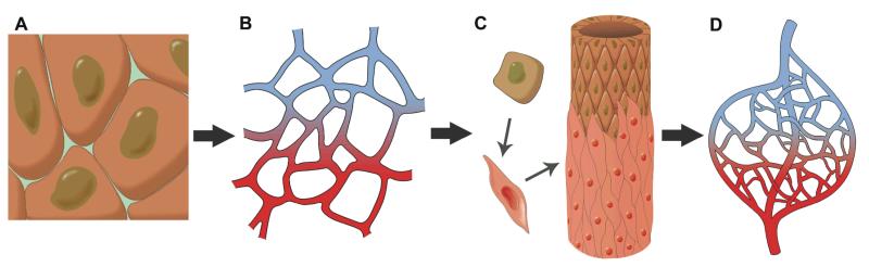
Endothelial progenitors (A) differentiate into arterial and venous endothelial cells that form a capillary plexus (B). The resulting vessels are sprouting and being stabilized by the recruitment of smooth muscle cells (C), to result in mature vasculature (D).

SEM images of collagen sponges for (A) PBS only treated scaffold (PBS), (B) Scaffold treated with EDC crosslinker in PBS without growth factors (PBS+EDC), (C) Scaffold with immobilized VEGF (VEGF), (D) Scaffold with immobilized Ang1 (Ang1), (E) Scaffold with lower dose of co-immobilized VEGF and Ang1 (1/2VEGF+1/2Ang1), and (F) Scaffold with higher dose of co-immobilized VEGF and Ang1 (VEGF+Ang1). Images at 100X with 500X insets. (G) Tensile modulus of scaffolds treated as above. # denotes statistically significant difference compared to PBS control. (P < 0.05; one-way ANOVA). Figure reproduced from [14] with permission. Copyright Elsevier.

Soluble VEGF (50ng/mL) and Ang1 (50ng/mL) were applied to cell-seeded collagen scaffolds treated with PBS alone (S-(VEGF+Ang1)), or scaffolds treated with EDC crosslinker in PBS (S-(VEGF+Ang1)+EDC). These scaffolds were compared to scaffolds with co-immobilized VEGF and Ang1 (VEGF+Ang1). 50,000 cells were seeded on the freshly made scaffolds. (A) XTT assay indicating final cell numbers in collagen scaffolds. (B) Lactate production rate. (C) Glucose consumption rate. * denotes statistically significant difference (P < 0.05; one-way ANOVA with post-hoc Tukey test). Figure reproduced from [14] with permission. Copyright Elsevier.
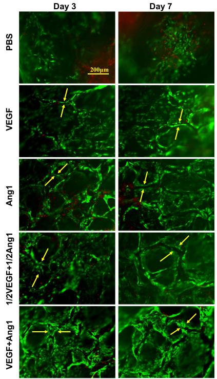
Representative live/dead images under fluorescence microscopy (173,333 cells initially seeded on the freshly made scaffolds; green represents CFDA staining of live cells and red represents propidium iodide (PI) staining of dead cells; tube is indicated between two yellow arrows). Figure reproduced from [14] with permission. Copyright Elsevier.
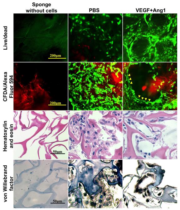
Images of PBS control and VEGF+Ang1 groups with live/dead staining (CFDA stains live cells green, PI stains dead cells red), cells (stained with CFDA) on collagen scaffolds labelled with Alexa Fluor 594 (red), hematoxylin and eosin staining, and von Willebrand factor staining. Sponges without cells are also shown. Figure reproduced from [59] with permission. Copyright Wiley.
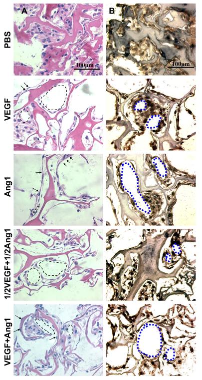
(A) Hematoxylin and eosin stained images (173,333 cells initially seeded on the freshly made scaffolds; arrows indicate elongated cells; arrowheads indicate circular structures). Note that darker pink in hematoxylin and eosin staining indicates the collagen scaffold, while the lighter pink is part of the cells. (B) Von Willebrand factor stained images (173,333 cells initially seeded; brown represents positive von Willebrand factor staining, blue represents counterstain; circular structures indicated by blue dotted outlines). Figure reproduced from [14] with permission. Copyright Elsevier.

Photographs of chicken eggs for (A) PBS and (B) VEGF+Ang1 groups. Arrow indicates location of collagen sponge. Figure reproduced from [59] with permission. Copyright Wiley.
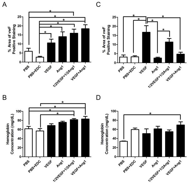
(A) Percentage of area with positive Factor VIII (FVIII) staining for Fresh scaffolds. (B) Hemoglobin concentration within Fresh scaffolds. (C) Percentage of area with positive FVIII staining for Aged scaffolds. (D) Hemoglobin concentration within Aged scaffolds. * denotes statistically significant difference (P < 0.05; one-way ANOVA with post-hoc Tukey test). Figure reproduced from [14] with permission. Copyright Elsevier.
References
Publication types
MeSH terms
Substances
Grants and funding
LinkOut - more resources
Full Text Sources
