

doi: 10.1002/jcp.22420.
RARγ is required for correct deposition and removal of Suz12 and H2A.Z in embryonic stem cells
Affiliations
- PMID: 20857416
- PMCID: PMC3369573
- DOI: 10.1002/jcp.22420
Item in Clipboard
RARγ is required for correct deposition and removal of Suz12 and H2A.Z in embryonic stem cells
J Cell Physiol.
2011 Feb.
Abstract
Retinoic acid (RA) induces embryonic stem cell differentiation. The effects of RA are mediated by retinoic acid receptors (RARs) that promote epigenetic changes controlling gene transcription. We show here that RARγ, in the absence of the ligand RA, is required for deposition of the histone variant H2A.Z and the polycomb group protein Suz12 at RA target genes, and that in embryonic stem cells both RARγ and Suz12 exist in a multi-protein complex in the absence of ligand. Addition of RA causes removal of H2A.Z and Suz12 from RARγ target genes when the genes are transcriptionally activated.
© 2010 Wiley-Liss, Inc.
Figures
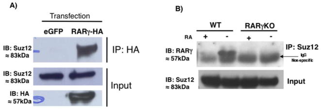
RARγ interacts with Suz12 in vivo. (A) Immunoprecipitation (IP) analysis of the interaction of RARγ with Suz12. COS cells transfected with eGFP or RARγ-HA expression vector. Cell extracts were IP using an anti-HA Ab and immunoblotted (IB) with an anti-Suz12 Ab (top panel). Whole-cell extracts were probed with anti-Suz12 to confirm equivalent Suz12 input before IP (middle panel) or anti-HA to check RARγ-HA expression (bottom panel). (B) ES cell extracts were IPd using Suz12 Ab and IBd with an anti-RARγ Ab (top panel). Whole-cell extracts were probed with the anti-Suz12 Ab to confirm equivalent Suz12 levels before IP (bottom panel). This experiment was performed three times.
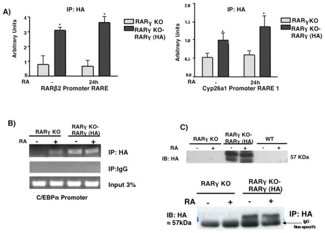
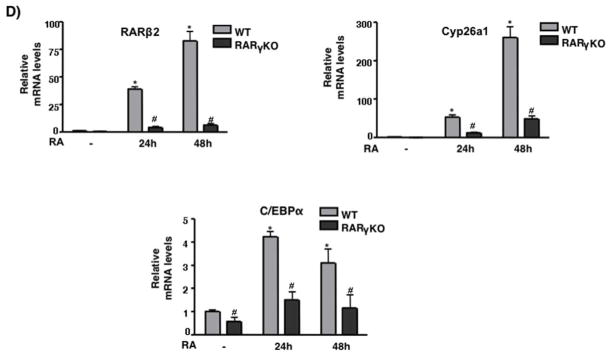
RARγ binding on RAR target genes in ES cells. ES RARγKO or RARγ-HA restoration cells were cultured +/− 1μM RA for 24h. (A) ChIP was performed using HA Ab or IgG and bound DNA was quantitated by real-time PCR. The data are presented as fold enrichment (mean ± S.E.) at RARβ2 or Cyp26a1 promoter. Error bars indicate standard error of three biological replicates corrected by GAPDH DNA bound for normalization. Statistically significant differences (p<0.05), asterisk. (B) Semi-quanitative PCR of RARγ-HA binding at the C/EBPα promoter. (C) Whole-cell extracts from RARγKO, RARγ-HA restoration, or WT ES cells were probed with the anti-HA Ab to confirm RARγ-HA expression. Representative Western blotting of three different biological replicates (top panel). IP analysis of RARγ-HA (bottom panel). ES cell extracts were IP’d using an Ab directed against HA and IB’d with an anti-HA Ab. (D) WT and RARγKO ES cells were cultured −/+ 1μM RA for 24h. Induction of the RA target gene mRNAs was measured by quantitative RT-PCR. Error bars, standard error of three biological replicates. Data in E were normalized to 36B4 mRNA. Statistically significant differences (p<0.05), asterisk (RA vs -) and # (WT vs RARγKO).
RARγ binding on RAR target genes in ES cells. ES RARγKO or RARγ-HA restoration cells were cultured +/− 1μM RA for 24h. (A) ChIP was performed using HA Ab or IgG and bound DNA was quantitated by real-time PCR. The data are presented as fold enrichment (mean ± S.E.) at RARβ2 or Cyp26a1 promoter. Error bars indicate standard error of three biological replicates corrected by GAPDH DNA bound for normalization. Statistically significant differences (p<0.05), asterisk. (B) Semi-quanitative PCR of RARγ-HA binding at the C/EBPα promoter. (C) Whole-cell extracts from RARγKO, RARγ-HA restoration, or WT ES cells were probed with the anti-HA Ab to confirm RARγ-HA expression. Representative Western blotting of three different biological replicates (top panel). IP analysis of RARγ-HA (bottom panel). ES cell extracts were IP’d using an Ab directed against HA and IB’d with an anti-HA Ab. (D) WT and RARγKO ES cells were cultured −/+ 1μM RA for 24h. Induction of the RA target gene mRNAs was measured by quantitative RT-PCR. Error bars, standard error of three biological replicates. Data in E were normalized to 36B4 mRNA. Statistically significant differences (p<0.05), asterisk (RA vs -) and # (WT vs RARγKO).
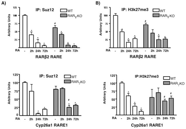
Suz12 and H3k27me3 proteins are present at the RARβ2 and Cyp26a1 RAREs in WT ES cells. WT and RARγKO ES cells were treated +/− 1μM RA for 2, 24 or 72h. ChIP was performed using anti-Suz12 (A) or anti-H3k27me3 Ab (B), and bound DNA was quantitated by real-time PCR. Each experiment was repeated at least three times. Data are show as % of input DNA before IP (mean ± S.E.) and normalized by GAPDH DNA bound. Error bars, standard error of three biological replicates. Statistically significant differences (p<0.05), asterisk (RA vs -) and # (WT vs RARγKO).
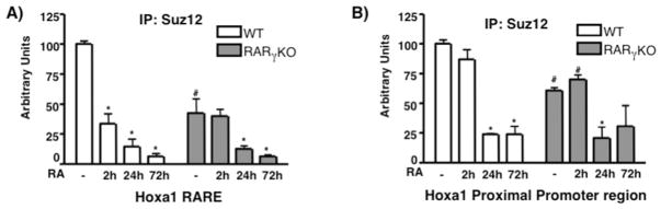
Suz12 is present at the 3′ downstream Hoxa1 RARE and at the proximal promoter region in WT ES cells. WT and RARγKO ES cells were treated +/− 1μM RA for 2, 24 or 72h. ChIP was performed using an anti-Suz12 Ab and bound DNA was quantitated by real time PCR at the RARE (A) or Hoxa1 PP region (B). Each experiment was repeated at least three times. Data are presented as %s of input DNA before IP (mean ± S.E.) and normalized by GAPDH DNA bound. Error bars, standard error of three biological replicates. Statistically significant differences (p<0.05), asterisk (RA vs -) and # (WT vs RARγ KO). 352×264mm (72 × 72 DPI)
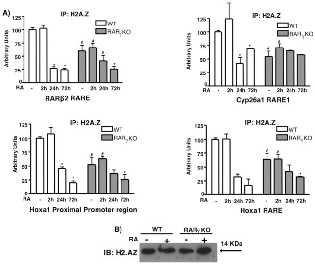
H2A.Z is present at RARγ target genes and its level decreases upon RA treatment of ES cells. ES WT and RARγKO cells were treated +/− 1μM RA for 2, 24 or 72h. ChIP was performed using an anti-H2A.Z Ab (A) and bound DNA was quantitated by real time PCR. Each experiment was repeated at least three times. Data are presented as %s of input DNA before IP (mean ± S.E.) and normalized to bound GAPDH DNA. Error bars, standard error of three biological replicates. Statistically significant differences (p<0.05), asterisk (RA vs -) and # (WT vs RARγKO). (B) Representative of three immunoblots showing H2A.Z protein levels in WT and in RARγKO cells.
References
-
- Barski A, Cuddapah S, Cui K, Roh TY, Schones DE, Wang Z, Wei G, Chepelev I, Zhao K. High-resolution profiling of histone methylations in the human genome. Cell. 2007;129(4):823–837. - PubMed
-
- Boyer LA, Plath K, Zeitlinger J, Brambrink T, Medeiros LA, Lee TI, Levine SS, Wernig M, Tajonar A, Ray MK, Bell GW, Otte AP, Vidal M, Gifford DK, Young RA, Jaenisch R. Polycomb complexes repress developmental regulators in murine embryonic stem cells. Nature. 2006;441(7091):349–353. - PubMed
-
- Campos EI, Reinberg D. Histones: annotating chromatin. Annu Rev Genet. 2009;43:559–599. - PubMed
-
- Chambon P. A decade of molecular biology of retinoic acid receptors. FASEB J. 1996;10(9):940–954. - PubMed
Publication types
MeSH terms
Substances
Grants and funding
LinkOut - more resources
Full Text Sources
