

Stromal stem cells from adipose tissue and bone marrow of age-matched female donors display distinct immunophenotypic profiles
- PMID: 20857424
- PMCID: PMC4340690
- DOI: 10.1002/jcp.22408
Stromal stem cells from adipose tissue and bone marrow of age-matched female donors display distinct immunophenotypic profiles
Abstract
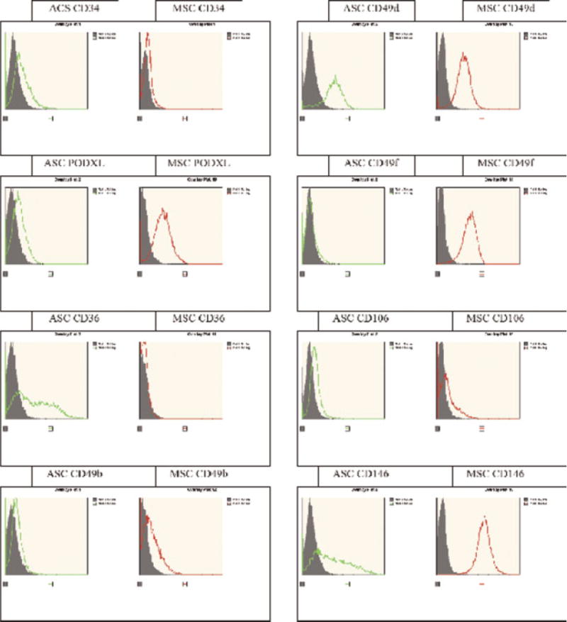
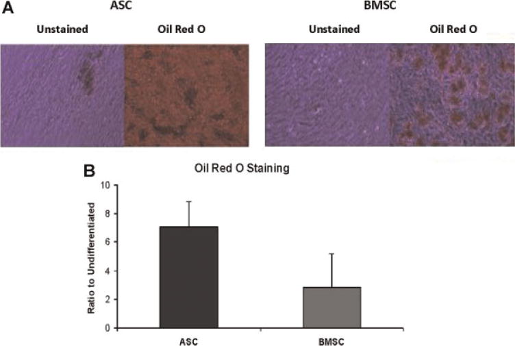
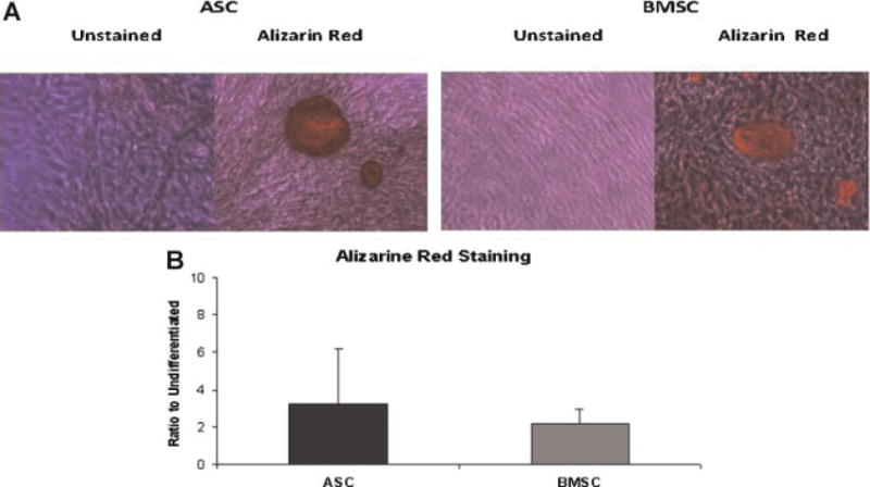
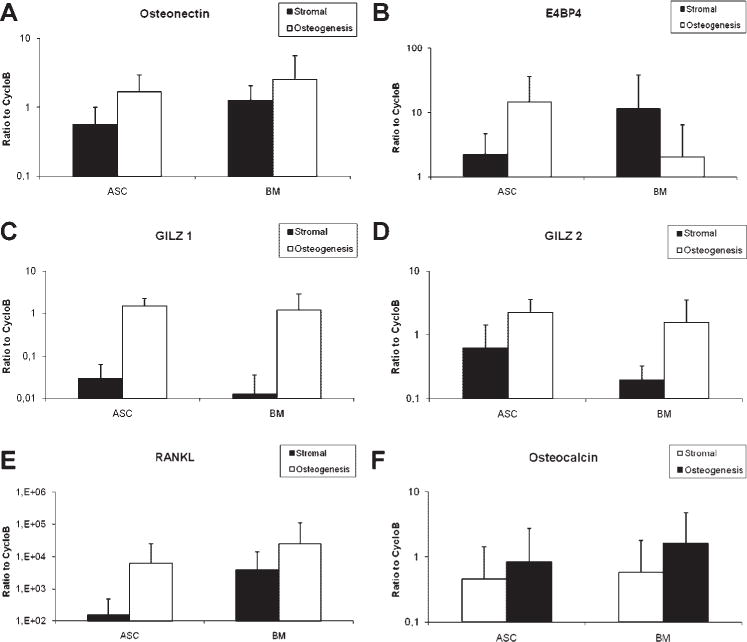
Adipose tissue is composed of lipid-filled mature adipocytes and a heterogeneous stromal vascular fraction (SVF) population of cells. Similarly, the bone marrow (BM) is composed of multiple cell types including adipocytes, hematopoietic, osteoprogenitor, and stromal cells necessary to support hematopoiesis. Both adipose and BM contain a population of mesenchymal stromal/stem cells with the potential to differentiate into multiple lineages, including adipogenic, chondrogenic, and osteogenic cells, depending on the culture conditions. In this study we have shown that human adipose-derived stem cells (ASCs) and bone marrow mesenchymal stem cells (BMSCs) populations display a common expression profile for many surface antigens, including CD29, CD49c, CD147, CD166, and HLA-abc. Nevertheless, significant differences were noted in the expression of CD34 and its related protein, PODXL, CD36, CD 49f, CD106, and CD146. Furthermore, ASCs displayed more pronounced adipogenic differentiation capability relative to BMSC based on Oil Red staining (7-fold vs. 2.85-fold induction). In contrast, no difference between the stem cell types was detected for osteogenic differentiation based on Alizarin Red staining. Analysis by RT-PCR demonstrated that both the ASC and BMSC differentiated adipocytes and osteoblast displayed a significant upregulation of lineage-specific mRNAs relative to the undifferentiated cell populations; no significant differences in fold mRNA induction was noted between ASCs and BMSCs. In conclusion, these results demonstrate human ASCs and BMSCs display distinct immunophenotypes based on surface positivity and expression intensity as well as differences in adipogenic differentiation. The findings support the use of both human ASCs and BMSCs for clinical regenerative medicine.
Copyright © 2010 Wiley-Liss, Inc.
Figures

References
-
- Abumrad NA, el-Maghrabi MR, Amri EZ, Lopez E, Grimaldi PA. Cloning of a rat adipocyte membrane protein implicated in binding or transport of long-chain fatty acids that is induced during preadipocyte differentiation. Homology with human CD36. J Biol Chem. 1993;268:17665–17668. - PubMed
-
- Aust L, Devlin B, Foster SJ, Halvorsen YD, Hicok K, du Laney T, Sen A, Willingmyre GD, Gimble JM. Yield of human adipose-derived adult stem cells from liposuction aspirates. Cytotherapy. 2004;6:7–14. - PubMed
-
- Bianco P, Riminucci M, Gronthos S, Robey PG. Bone marrow stromal stem cells: Nature, biology, and potential applications. Stem Cells. 2001;19:180–192. - PubMed
-
- Bunnell BA, Estes BT, Guilak F, Gimble JM. Differentiation of adipose stem cells. Methods Mol Biol. 2008b;456:155–171. - PubMed
Publication types
MeSH terms
Substances
Grants and funding
LinkOut - more resources
Full Text Sources
Other Literature Sources
Medical
Research Materials
Miscellaneous
