

doi: 10.1002/anie.201003761.
Readily accessible bicyclononynes for bioorthogonal labeling and three-dimensional imaging of living cells
Affiliations
- PMID: 20857472
- PMCID: PMC3021724
- DOI: 10.1002/anie.201003761
Item in Clipboard
Readily accessible bicyclononynes for bioorthogonal labeling and three-dimensional imaging of living cells
Angew Chem Int Ed Engl.
.
No abstract available
Figures
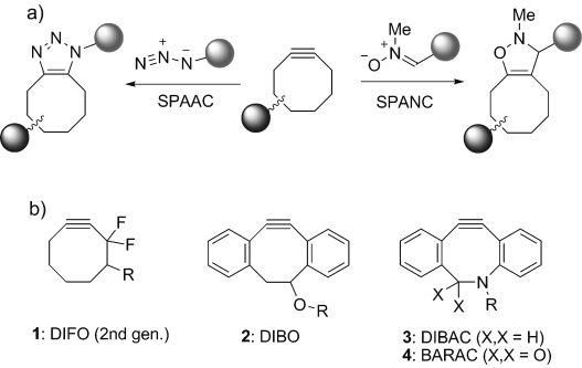
Reactions and structures of cyclooctyne compounds for strain-promoted cycloaddition. a) Cycloaddition with azide (SPAAC) or nitrone (SPANC). b) Structures of the most commonly employed cyclooctynes.
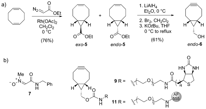
a) Synthesis of 9-hydroxymethylbicyclo[6.1.0]nonyne (endo-6). b) Structures of nitrone 7 and BCN conjugated to biotin (9) or Alexa Fluor 555 (11). THF=tetrahydrofuran.
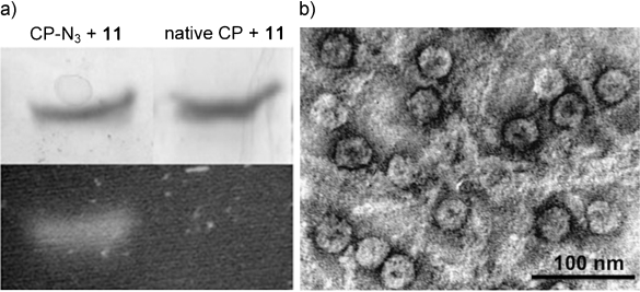
BCN modification of azido-containing virus capsid protein and assembly into virus capsids. a) SDS-PAGE analysis of reaction of BCN-AF555 conjugate (11) with capsid protein containing azide (left) or without azide (right). Top: Coomassie Brilliant Blue staining, bottom: fluorescence image. b) TEM pictures of fluorescent capsids (images recorded on a JEOL 1010 TEM, the sample was deposited on a hydrophilized Formvar carbon-coated TEM grid and consequently negatively stained with 0.2 % uranyl acetate).
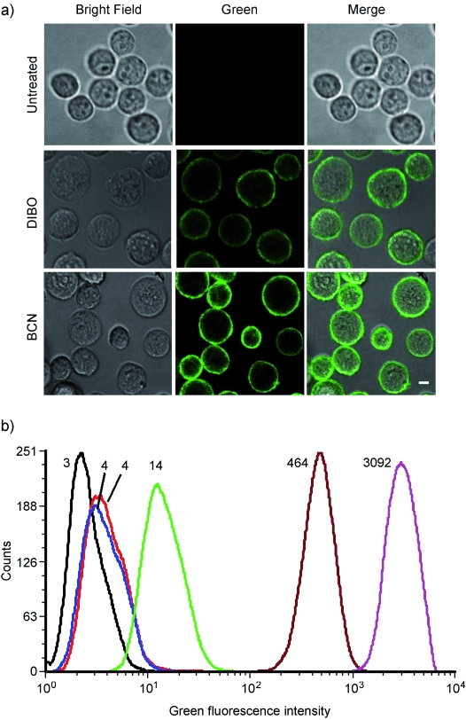
Surface and total fluorescence intensity of MV3 melanoma, cultured in the absence or presence of Ac4ManNAz (50 μm ), followed by labeling with a cyclooctyne-biotin conjugate and secondary labeling with streptavidin-Alexa Fluor 488. a) Representative confocal images of unlabeled cells (top), cells labeled with DIBO-biotin (middle) or BCN-biotin 9 (bottom). Bar: 10 μm. b) Label intensity assessed by flow cytometry, indicated as mean fluorescence intensity (MFI). Numbers denote the average of green fluorescent cells for that particular experiment. Black trace: untreated; red trace: Ac4ManNAz + SA-AF488; blue trace: w/o Ac4ManNAz + DIBO + SA-AF488; dark red trace: Ac4ManNAz + DIBO + SA-AF488; green trace: w/o Ac4ManNAz + BCN + SA-AF488; magenta trace: Ac4ManNAz + BCN + SA-AF488.
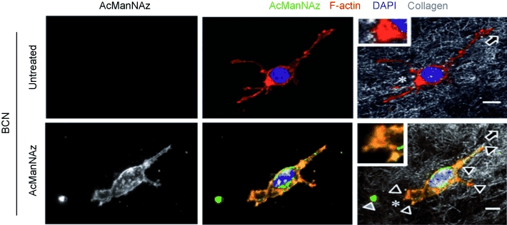
Live-cell staining and redistribution of glycans during invasive cell migration through a three-dimensional collagen matrix. Focal accumulation of sialic acid on migrating MV3 melanoma cell at interaction sites to collagen fibers and partial colocalization with F-actin. Insets, trailing edge. Direction of migration was determined from retraction fibers (asterisks) and deposited sialic acid-rich material lacking F-actin from the cell rear (gray arrowhead).[28] Bar: 5 μm. Focalized glycan distribution at cell matrix interactions (black arrowheads).
References
Publication types
MeSH terms
Substances
LinkOut - more resources
Full Text Sources
Other Literature Sources
Medical
